Effect of growth factors on chondrogenic differentiation of rabbit mesenchymal cells embedded in injectable hydrogels
Keun Hong Park1 and Kun Na 2*
College of Medicine, Pochon CHA University, Cell and Gene Therapy Research Institute, 606-16 Yeoksam 1-dong,
Kangnam-gu, Seoul 135-081, Korea1 and Department of Biotechnology, The Catholic University
of Korea, 43-1 Yokkok2-dong, Wonmi-gu, Bucheon, 420-743, Korea 2
JOURNAL OF BI OSCI ENCE AND BI OENGI NE ERING © 2008, The Society for Biotechnology, Japan
Vol. 106, No. 1, 74–79. 2008
DOI: 10.1263/jbb.106.74
Summary: The prospect of generating hydrogels with implanted growth factors to aid in tissue regeneration and healing is an alluring area of research. The three-dimensional scaffold that a hydrogel produces allows for increased cell differentiation, proliferation, and engraftment in the targeted area. Additionally, the degradation rate, biocompatibility, and mechanical properties of such an implant can be manipulated to suit a vast array of purposes. In this study, a hydrogel scaffold of poly(NiPAAm-co-AAc) and transforming growth factor beta-3 (TGF β-3), or basic fibroblast growth factor (bFGF), or not factors was embedded with rabbit mesenchymal cells. The polymer was then implanted into mice. The implant was then removed after 1, 4, and 8 weeks and various measurements were taken.
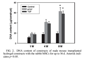
Results: Total DNA content of the hydrogel was calculated and tabulated as a function of time in figure 2. 
Here it is clearly seen that the amount of DNA and correspondingly the number of cells that proliferated are highest for the hydrogels with growth factors implanted. All of the samples needed time to grow. The gels implanted with bFGF and TGF β-3 increased 300% and 280% from week 1 to week 8. Additionally, the tissue that was formed was denser.
Glycosaminoglycans (GAG) are a cartilage component whose presence and proliferation were used to measure ECM integration. Consistent with the proliferation of cells, cells subjected to growth factors showed a much higher amount of GAG. The composition of GAG was also favorable in the growth factor embedded samples. Both bFGF and TGF β-3 showed higher total collagen content in the hydrogel scaffold, and TGF β-3 showed an additional increase in type II collagen. The group then used RT-PCR to visualize gene expression and found that the scaffold containing TGF β-3 had an increased type II collagen mRNA and a decreased expression of type I collagen, an osteogenic factor. Safranin O staining revealed that cells in the growth factor embedded hydrogel were rich in ECM components, while those in a hydrogel lacking the factors only had ECM in the immediate vicinity of the cell.
Critique: The group did a good job of showing the effect of using TGF β-3 for increased chondrogenic differentiation. However, the scope of the study was very limited. For future studies, it would be useful to include information about different concentrations of TGF β-3 that optimize growth, as well as a variety of hydrogel formulations. Additionally, the way in which TGF β-3 is embedded in the hydrogel scaffolding could have profound effects on the integrated cells.

8 comments:
Do they ever correlate the rate at which TGF-B3 and bFGF diffuse out of the hydrogel? It would be important to know how the rates that these factors diffuse into the site of interest affect cell proliferation.
You mention: "Here it is clearly seen that the amount of DNA and correspondingly the number of cells that proliferated are highest for the hydrogels with growth factors implanted. All of the samples needed time to grow. The gels implanted with bFGF and TGF β-3 increased 300% and 280% from week 1 to week 8."
I am a bit confused on how they measure proliferation. It seems that Figure 2 only shows increased levels of DNA - is that generally accepted to correlated to cell proliferation? Or did they do another assay/measurement to determine proliferation?
If not, then it would be worth while to do such an assay, as it does not intuitively make sense DNA levels necessarily correspond to cell proliferation.
On a separate note, I like your future works suggestions and how you gave enough background to understand the implications of the paper.
I was really disappointed in how this paper really limited its scope. The do show that cells proliferated on the hydrogel, but it would have been interesting if they could really narrow down what it was in the hydrogel that allowed the proliferation. I'm certain the mechanical properties of the gel, such as stiffness would also be really helpful in understanding the cell growth.
Also, if they could use some sort of interpenetrating polymer network, they could be able to control the mechanical properties of the gel and the actual presence of growth factors.
I guess this is more of a comment on the paper itself than your summary, which I thought did a good job of summing up the paper.
I understand that a hydrogel is a cross-linked system which is mainly composed of water, where the jelly-like material can range from a "softer" gel to a "harder" gel. Did this paper talk about the strength of the gel and its role in the effect of growth factors on chondrogenic differentiation?
From the paper, it appears that the researchers embedded the growth factors (GFs) into the hydrogel by putting the gel in an aqueous solution containing the GF. Is this how GFs are usually embedded into hydrogels? I think you can also chemically bond the GF to the hydrogel. For future studies, the researchers could use different methods of incorporating the GF and compare the results.
Raj: The paper said they measured specifically the amount of double-stranded DNA. I assume this could give an accurate account of cell proliferation because the only dsDNA would be genomic DNA which all the cells have the same amount of.
Is the addition of the growth factors the only difference between the control group and the hydrogel + growth factors group? If not, then perhaps some other reagent could account for the collagen and GAG growth. In the future, it would be interesting to investigate whether the growth factors are specific to a type of collagen growth and also how the design of the hydrogel influences cell proliferation and cell differentiation. It is possible that designing a hydrogel that is more similar to the natural environment of cell tissues result in greater cell proliferation/cell differentiation.
Derek: Nope, I don't think they ever do that but yes it would be a good factor to take into account.
Raj: They determined cell number by staining double stranded DNA and measuring it using a PicoGreen assay. I'm not particularly familiar with the assay but they seem pretty confident in its ability to estimate cell number.
Ashin: I can't really speak for them so I'll just nod. I think they were mostly experimenting with growth factors and the hydrogel was just there to distribute the chemicals and act as a matrix for cell growth. But certainly it would have an effect on cell growth and further studies would have to be done to characterize these happenings.
Neeraj: Similar to above, I think they were just testing growth factors, but varied compositions of hydrogels would be needed to see the effects and that would be interesting to find out.
Mimi: I'm not sure how GF are normally attached but it is an interesting question. Also, thanks, you got my back.
Karthik: I think the only difference was the presence of GF.
I think your suggestions for future studies are very valid. I'm surprised they didn't do parellel studies when different concetrations of the growth factors were used. It would be very valuable data. It would also be good to know where the implants are put inside the animals to characterize where growth is the fastest/slowest, etc.
Post a Comment