Saturday, October 31, 2009

Cell Lysis and Protein Extraction in a Microfluidic Device with Detection by a Fluorogenic Enzyme Assay

Eric A. Schilling, Andrew Evan Kamholz and, Paul Yager. Analytical Chemistry 2002 74 (8), 1798-180.

Summary:

A novel Mylar microfluidic device for lysis of bacterial cells and detection of b-galactosidase was developed and described. The chemical-based lysis device consisted of an H-filter with three inlets and two outlets (Figure 1). A suspension of E. coli (~108 cells/mL) and the lytic agent were pumped in separately and met in the main lysis channel, which depends on diffusion of the detergent to lyse cells. Bacterial Protein Extraction Reagent (BPER) was chosen as the lytic agent in order to permeabilize the bacterial cell membranes and allow release of b-galactosidase without affecting the catalytic activity of the enzyme. The lysis channel split into two channels downstream, a controlled outlet and a detection channel. The geometry of the device allowed cells to be discarded out of the outlet, while smaller molecules had sufficient time to diffuse into the side of the channel that split off into the detection channel. A fluorogenic assay downstream of the lysis allowed for detection of b-galactosidase. b-D-galactopyranoside (RBG) was pumped into the third outlet, which flowed into the detection channel to allow for fluorescent detection of b-galactosidase. b-gal catalyzed the reaction of RBG into resorufin and D-galactose. Resorufin fluoresces when excited at 570 nm and can be detected at 594 nm. A numerical model of the system allowed the researchers to determine the concentration of b-gal in the lysate by integrating Michaelis-Menten kinetics and diffusion coefficients of b-gal, RBG, and resorufin.

Before experimenting with the device, the researchers conducted 3D modeling using CoventorWare to create a velocity profile for b-gal (Figure 2). The researchers assumed that b-gal flowed into the cell inlet, rather than modeling the time taken for lysis to occur. They stated that this error is negligible because previous experiments demonstrate a lysis time of several seconds, which is two orders of magnitude less than the residence time b-gal is present in the channel, 190 seconds.

The researchers also ensured that no cells entered the detection channel by taking dark-field images of the functional device within seconds of fluorescent images. The fluorescent images displayed resorufin fluorescing while the dark-field images showed no cells. In order to back up these findings, the researchers also introduced a fluorescent DNA stain (SYTO 9) to the cells before flowing them into the device and used fluorescent imaging to detect resorufin (red) without SYTO 9 (green) in the detection channel, and both present in the cell outlet.

Significance:

The paper states that the ability to integrate cell lysis into microfluidic chips already developed for analysis would increase the portability and ease of cellular analysis. Analytical techniques such as immunoassays, PCR amplification, and DNA analysis are some of the possible downstream uses of an integrated lysis-analysis microfluidic chip. Several other on-chip devices for lysis have been developed, but these require an external power source and the researchers expressed concerns about the cost of such devices. The researchers feel that their microfluidic device will be both easier to use and fabricate and less expensive to manufacture than these other devices. Although the researchers have thus far only worked with E. coli cells, they state that the dimensions of the device as well as the reagents used could be easily changed in order to lyse and analyze a wide range of cell types. Also, although b-gal is the only molecule detected in the experiments, the researchers have stated that changes in flow rates will allow for smaller molecules to be fractioned out of the cell stream. They chose b-gal because it was a relatively large molecule and could be detected with a simple fluorogenic assay.

Critique:

The researchers do a good job of explaining their choices of reagents, flow rates, and geometry and sufficiently describe controls and experimental checks, but they do not address the feasibility of using the device for further analysis. According to the papers, b-gal has the ability to diffuse almost 100um from the place it was originally released from the cell during the 190 seconds that it resides in the lysis channel. With a channel of 1000m wide and 100um deep, it seems as though a great deal of b-gal would remain in the left half of the channel. This b-gal would be discarded with the cells through the controlled outlet. Therefore, the amount of b-gal harvested by the microfluidic device is much less than the amount initially released by the cells. In fact, according to the paper, only 10% of the b-gal present in the cell stream actually flowed into the detection channel. Although the researchers have included information about the b-gal concentration in the detection channel, there is no discussion about the amount needed for downstream analytical techniques. This paper is a proof-of-concept that E. coli cells can be lysed on a microfluidic chip using chemical lysis, but it does not address the feasibility of using the lysate for later analysis. Figures such as amount of enzyme harvested from the final outlet versus number of cells pumped through the device should be included in future work.

Activation of Na+/H+ and K+/H+ exchange by calyculin A in Amphiuma tridactylum red blood cells: implications for the control of volume-induced ion flu

Citation:

Alejandro Ortiz-Acevedo, Robert R. Rigor, Hector M. Maldonado, and Peter M. Cala

Activation of Na+/H+ and K+/H+ exchange by calyculin A in Amphiuma tridactylum red blood cells: implications for the control of volume-induced ion flux activity
Am J Physiol Cell Physiol 295: C1316-C1325, 2008. First published doi:10.1152/ajpcell.00160.2008


Summary:

In this paper, the authors sought to study the mechanism behind the Na+/H+ and K+/H+ exchange that is crucial to cell volume maintenance. Cells regulate their volume when placed in hypo- or hyper- osmotic environments through the efflux of K+ and Cl- (when swelled) and the influx of Na+ and Cl- (when shrunk). These channels have been shown to be affected by phosphorylation and dephosphorylation cascades, but the mechanism is not understood. Based on previous studies, the authors hypothesize a phosphorylation-based scheme for the regulation of intercellular volume, and test this mechanism through a variety of conditions. These experiments are based on the inhibitory properties of calyculin A (CLA), ouabain, and 5-(N-ethyl-N-isopropyl)-amiloride (EIPA). CLA is a phosphotase inhibitor, so introduction into culture will result in up-regulation of phosphorylation cascades. Ouabain is a Na+/K+ ATPase inhibitor, and is used to eliminate alternative sources of ion flux. EIPA is an inhibitor of both the Na+/H+ antiport and potentially of the K+/H+ antiport. Together, these three molecules are used to regulate ion flux into and out of a cell during osmotic stresses.


The effect of CLA on cells in isotonic solutions was measured by measuring the flux of radio-labeled ions in solution. Even without osmotic stresses, cells exposed to CLA demonstrated a net Na+ uptake and a net K+ loss (Figure 1). This is interesting, because upregulating phosphorylation will induce cell responses to both swelling and shrinking simultaneously. This suggests that both pathways are either controlled by a common regulatory event or sequentially linked in some way. To test the latter scenario, normal K+/H+ and Na+/H+ channel function was blocked by using ‘null media’ (media designed to maintain those channels at a thermodynamic valley to eliminate driving force for ion flow). When cells in this media were exposed to CLA, they continued to exhibit ion flux, which suggests that this flow is not secondary to another channel. Next, EIPA, a Na+/H+ Exchanger 1 (NHE1) inhibitor, was used to block access to Na+/H+ exchangers in the cell membrane, while also blocking homologous NHE’s that mediate K+/H+ exchange as well. Blocking of ion channels resulted in significantly decreased ion flux activity, even when cells were exposed to CLA (Figure 6). This further suggests that the mechanism for cell volume control runs through phosphorylation of K+/H+ and Na+/H+ channels.


The authors next seek to determine if both the cell swelling and cell shrinking responses are controlled by the same pathway. They propose that a shrinkage-activated kinase activates Na+/H+ exchange but is suppressed by swollen cells, and vice versa. To test this, a new experimental design was created to zero out phosphotase activity prior to exposure to osmotic stresses. The ‘null media’ was used again to ensure that no ion flow occurred while cells were incubated with CLA. After this incubation, cells are exposed to media without CLA and ion flow is measured. When an isotonic solution was used for this experiment, cells experienced no net ion flow during the incubation period, and a subsequent net influx of Na+ and efflux of K+ (Figure 7). However, when cells were incubated in a hypotonic solution, they experienced primarily a K+ efflux after exposure to non-‘null media’. Likewise, cells in a hypertonic solution experienced primarily a Na+ influx after media change (Figure 8). This result suggests that the pathways for each ion channel flux are independent of the other, but can be regulated at a common step by a CLA-sensitive phosphotase.


Significance:

This paper proposes and tests a novel mechanism for the control of cell volume through sodium and potassium channels. While prior work had already hinted at the link between phosphorylation and regulation of ion channels, this paper creates a model that explains selective ion flow through two independent pathways (one for cell swelling, one for cell shrinking) that share a common regulator step (for isotonic solutions).


Critique:

There were some potential systemic issues with their protocol that might adversely affect data validity. Throughout the experiments, rabbit red blood cells were held at 23ºC, which is significantly lower than body temperature (37ºC). The effect the lowered temperature may have on membrane permeability is unknown, and may skew results unpredictably. All ion flow measurements were taken using radioisotopes of ions and a gamma counter or beta scintillation counter. The authors described how they would cut the tube “just above the cell pellet to minimize contamination by extracellular isotope” during readings. However, this seems very inconsistent and irreproducible, since with varying sized pellets would come varying sized cuts, and the background radiation would be impossible to blank properly.


Additionally, I was not entirely convinced by their model for how the two pathways are related, while at the same time controlled upstream by a shared phosphotase. While their proposal seems entirely reasonable, the data do not conclusively point to this mechanism as the best or only one. While the experiments conducted have shown that the proposed mechanism is possible, I feel that further tests would need to be conducted to rule out alternative mechanisms. One particular area of interest is in the possibility that alternative ion channels played a part in this mechanism. While the authors tried to control for channels such as the Na+/K+ ATPase and the Na+/H+ or K+/H+ exchangers, it seems likely that there were additional ion channels that should have been accounted for. Overall, while this paper provides a lot of evidence supporting the author’s proposed mechanism, it does not provide evidence sufficiently disproving the possibility of alternative mechanisms.


Figures:


Friday, October 30, 2009

Dynamics of Endocytosis and Exocytosis of Poly(D,L-Lactide-co-Glycolide) Nanoparticles in Vascular Smooth Muscle Cells

Summary:

The formulation of macromolecular agents such as proteins, oligonucleotides, and DNA in drug delivery systems has drawn interest because of their effectiveness in treating diseases. However, the effectiveness of such systems is severely limited because the macromolecules often need to enter cells in order to access their sites of action. They are hindered by inefficient uptake and their susceptibility to degradation during an endocytosis based uptake process. While other systems such as viruses and protein transduction domains have been used to successfully enter cells, they are able to carry only a few types of therapeutics. Moreover, some of these systems are limited by toxicity and immunogenicity concerns.

The authors of this paper have previously shown that biodegradable poly-PLGA nanoparticles can escape degradation and deliver therapeutics into the cytoplasm. They are also able to release the agent in a sustained manner. However, their internalization and retention inside the cell has not been studied in detail. Therefore, in this paper, the authors characterize endocytosis and exocytosis of PLGA nanoparticles in human arterial vascular smooth muscles cells (VSMCs).

Dose dependent and time dependent cellular uptake of nanoparticles were studied along with the process of exocytosis under normal and metabolically inhibited conditions. BSA was used as a macromolecule model and 6-coumarin as a fluorescent marker. These were loaded onto the nanoparticles using a double emulsion-solvent evaporation technique. The dye was shown to stay within the nanoparticle throughout the intake and study process. Cells were plated with two different media: regular growth medium and serum-free medium, both containing nanoparticles. In order to study dose-dependent nanoparticle uptake, the cells were incubated with different concentrations of the nanoparticle suspension for 1 hour. To study time-dependent nanoparticle uptake, cells were incubated at a given nanoparticle concentration for different time periods. Exocytosis under normal conditions was studied by initially incubating the cells with nanoparticles for a given time period in the two media types. The uninternalized nanoparticles were then washed off and the cells were incubated in growth medium without nanoparticles. Cells were removed and lysed at varying time intervals to determine the fraction of nanoparticles retained. In order to study the metabolic inhibition of nanoparticle exocytosis, cells were first incubated with nanoparticles for a given time period, then washed and incubated in regular medium containing sodium azide and deoxyglucose. These molecules inhibit the production of ATP and thus reduce the ability of a cell to perform exocytosis. Again, cell lysates were studied to determine nanoparticle concentration. Finally, Lucifer yellow, a fluid phase marker was used as a control for exocytosis. Cell lysates were processed to determine the nanoparticle levels through high-performance liquid chromatography. These processes were also visualized using confocal laser scanning microscopy.

The uptake of nanoparticles was shown to be both dose and time dependent. In the dose-dependent study, uptake was linear at lower doses (10-100g), tapering off at higher doses (500-1000g). The results of the time-dependent study showed that uptake began as early as 10-30 seconds (Figure 3), was rapid during the first two hours and reached saturation in 4-6 hours. Figure 2 displays a graph of the uptake. The exocytosis study showed that a majority (65%) of the internalized nanoparticles were exocytosed immediately after the removal of nanoparticles from the external growth media. Furthermore, exocytosis was inhibited in the presence of metabolic inhibitors, confirming that endocytosis was the mode of uptake. Interestingly, the exocytosis was almost completely inhibited when cells were places in serum-free media whereas the addition of BSA to the media induced exocytosis. The authors suggest that this indicated the protein is either adsorbed or carried along with the nanoparticles into the cells and interact with the exocytic pathway, leading to the increased exocytosis.

Conclusion:

Overall, the study shows that the nanoparticles are internalized rapidly in a saturable, energy-dependent process. However, a majority of the internalized particles are exocytosed immediately when the concentration gradient across the cell membrane is removed. Nonetheless, the authors speculate that this may not be a problem in vivo because the concentration of nanoparticles will not fall as rapidly in the extracellular matrix. This would allow the cells to reach a mass transport equilibrium and thus retain more nanoparticles. Among other things, the mechanisms of endocytosis and exocytosis and the effect of nanoparticle formulation and composition on the processes need to be explored before further conclusions can be drawn.


Thursday, October 29, 2009

Genetically Engineered Nanofiber-Like Viruses For Tissue Regenerating Materials

Genetically Engineered Nanofiber-Like Viruses For Tissue Regenerating Materials

Anna Merzlyak, Shyam Indrakanti, and Seung-Wuk Lee*

Department of Bioengineering, UniVersity of California, Berkeley, Physical Biosciences DiVision, Lawrence Berkeley National Laboratory, Berkeley Nanoscience and Nanoengineering Institute, Berkeley, California 94720

Nano Letters 2009 Vol 9, No 2 846-852

This paper introduces a novel biomaterial composed of nanofiber-like viruses, M13 Bacteria Phage. These viruses are capable of providing an ordered extracellular matrix for cell adhesion and display signaling motif on their coat protein to direct cell behavior and growth. As shown in Figure 1, The different functionalities of viruses come from their shape, charge, peptide displayed which can be modified through chemical or genetic approaches.


RGD and IKVAV peptides were displayed at the N-terminus of viral major coat protein VIII (pVIII). RGD is a cell adhesion integrin binding motif and IKVAV is a laminin motif known to promote neural adhesion and extention. The pVIIIs are expressed 2700 times on a single phage, representing 99% of the phage surface. Peptides of up to 8 amino acids can be inserted at pVIII.

Non-toxicity of the phages was shown through CyQuant assay. CyQuant assay is based on fluorescent intensity after cyanine dye binds to nucleic acid, which is proportional to cell count. Cell culture with phage had similar CyQuant fluorescent readings in comparison to the Cells with out phage on day 1, 3 and 5.

To verified the effect of RGD and IKVAV, hippocampal neural progenitor cells (NPCs) were selected for the potential application of neural regeneration in case of spinal cord injury. Immunostainning showed RGD and IKVAV phage colocalize with the cell while the wild phage does not. This shows the specificity interaction of their engineered phage with Cell. Further microscopy studies from SEM showed NPCs on RGD or IKVAV spread along the direction of the phage film and well spread while the cell on wild phage showed aggregation instead of extension.

3D Phage fiber in agrose gel was then studied. RGD and IKVAV phage matrices were able to promote unidirectional cell spreading and elongation by 30% compare to wild phage. These results shows the phages were chemically encouraging and guides the cell to grow in specific direction. This is particular useful in neural regeneration, because neural network is well known for its directionality that provide the platform for our cognitive functions. The ability to recreate such directional guidance is significant.

Potential Improvement:

During 3D culture experiments, RGD, IKVAV, and Wild phages were compared. I will add a Laminin positive control to show how good the RGD and IKVAV peptides compare with the native Laminin matrix that NPCs grow on in vivo.

The other improvement might be the colocalization of the phage with the cell. Author suggested specific interaction. But colocalization does not mean interaction. She did not go the extra step to prove it is actually interaction. I will do an additional binding assay to quantify the interaction.

Hematopoietic Stem Cell Development Is Dependent on Blood Flow

Hematopoietic Stem Cell Development Is Dependent on Blood Flow. Trista E. North, Wolfram Goessling, Marian Peeters, Pulin Li, Craig Ceol, Allegra M. Lord, Gerhard J. Weber, James Harris, Claire C. Cutting, Paul Huang, Elaine Dzierzak, and Leonard I. Zon. Cell. May 2009; (137)4: 736-748.

Available at: http://www.sciencedirect.com/science?_ob=ArticleURL&_udi=B6WSN-4W9297H-K&_user=4420&_rdoc=1&_fmt=&_orig=search&_sort=d&_docanchor=&view=c&_acct=C000059607&_version=1&_urlVersion=0&_userid=4420&md5=5be731494083d83e4456767fea553c35

It is important to look at the development of hematopoietic embryonic stem cells because the regulation of development is very similar to that of the maintenance and recovery for adult hematopoietic stem cells (HSC). As such, learning the development of HSCs can give insight into potential treatments for aging and damaged tissue. One particularly interesting location of study is the aorta-gonads-mesonephros (AGM) region of a heart during embryogenesis due to the fact that this region is densely seeded with hematopoietic stem cells. This paper uses the AGM region as a model to test the hypothesis that mechano-transduction from pulsatile blood flow helps HSCs develop during embryogenesis. In addition this paper attempts to elucidate key pathway intermediates in order to identify which pathways are being affected by mechano-transduction in order to promote HSC development.

To begin with, this paper attempts to establish that HSC development is enhanced by pulsatile blood flow. The reason for such suspicion is that during early development, the hearts of vertebrates starts to pump even though they can easily rely on diffusion for oxygenation. One possible explanation is the heart starts to pump in order to promote HSC cell development in the AGM. In order to test this theory, the researchers used a mutant zebra fish called silent heart (sih) in which the heart does not beat during embryogenesis. In the absence of blood flow during embryogenesis, there is less development. Development was measured in number of HSCs present, inferring that a higher number means superior development.

Initially, the researchers tried to determine what factors/ signaling pathways were at play. First they wanted to confirm that regulators can affect development. Drawing from knowledge from literature, they postulated that factors runx1/cmyb+ are directly related to HSC development. They tried various inhibitors and enhancers to determine what key intermediates are affecting the runx1/cmyb+ concentration. After trying a menagerie of inhibitors and enhancers, they elucidated a lot of effectors such as: digoxine, L-NAME, etc. Looking at aorta diameter, HSC count and runx1/cmyb+ levels, they concluded there are many factors that can contribute to development including nitric oxide (NO).

Next, they inferred that if in fact mechano-transduction is lowering some regulatory pathway in order to slow down HSCs growth, then one can “rescue” these cells with no mechano-transduction by fixing the regulation. The researchers determined that it was NO that was the major contributor in HSC development because NO was able to rescue zebra fish development in the absence of pulsatile flow. They also reconfirmed this by knocking out the downstream pathway for NO signaling and confirming that this has the same effect as knocking out NO.

Next, the researchers established that NO signaling affects development prior to mechano-transduction. They tested this by adding in a NO inhibitor (L-NAME) and seeing impeded growth. The researchers also wanted to see if the effect of NO signaling was cell autonomous. They achieved this by transplanting NO treated cells into a blastula and watching only the transplanted cells develop. They distinguished transplanted cells from their neighbors by a fluorescent marker. As well, they were interested in other pathways talking with NO signaling, namely the wnt and notch pathways. They used a mindbomb mutant (mb) in which the notch pathways was greatly knocked down. This showed a greatly reduced hematopoietic stem cell development. They also had a constitutively on notch mutant in which reduced HSCs showed increase development and they were able to knock down this development by adding a NO inhibitor, L-NAME. They confirm from this that notch is upstream of NO. The same scheme was used on the wnt pathway.

For completeness, they attempted to look at other pathways developed by blood flow. In order to search for these pathways, they look for phylogenetic trees and genomic similarities in order to determine similar pathway intermediates. They found nos1 and nos3, both NO synthases in other cell types. Reduction of NO resulted in reduced development in those cell types.

Lastly, they confirmed that what they saw in zebra fish is conserved in mice, by taking using the L-NAME inhibitor and using it in pregnant mice and doing histological studies to determine reduced development of hematopoietic stem cells.

Discussion and Criticism

One improvement (nice way of calling something wrong) to this paper is that in the cell autonomy part of the paper. The paper claims that the effect of nos1 (which is supposed to reduce HSC growth) is not context dependent (not affected by neighboring cells.) To show this is autonomous of the context the cells are in, they took nos1 knockdown mutants and implanted that into a blastula of healthy cells. Tracking of these cells was done with a fluorescent GFP tag. The one criticism to this is that while the experiment does suggest that this is context dependent it is not completely conclusive. The problem is that you are transplanting an ensemble of nos1 knockdowns into the blastula. Unfortunately, by transplanting a group of cells, you may be transplanting an environment. It is possible that the small isolated colony of cells may regulate themselves such that they do not develop. What I would do to be more conclusive is that I would want to transplant a single cell into the new environment to check for cell autonomy.

Another potential improvement is in the interacting signaling pathways. It is very difficult to assess the behavior of a system of pathways from a simple trial and error test. The paper identifies that both notch and wnt pathways positively affects HSCs by increasing NOS. However, this assumes that the relationships are linear. This is probably not the case as it can be very possible that wnt and notch both, by themselves, increases development, but together, they it will reduce HSC development. Furthermore, it is suggested that increased NO is the only factor. This might in fact be an ensemble effect. To improve this into more meaning and predictive information, I would construct consult a systems biologist in order to construct a web or pathway instead of single interaction. Then I would test interacts based on possible topologies to confirm and conclude a model of signal transduction.

As far as big picture problems go, I would do more tests to confirm the relevance of these findings in developmental systems in mature systems. The end goal of these experiments is to help with tissue repair in adult systems. As such, this information would only be useful if it does in fact pertain to adult systems. As such, I would run experiments in mature cells to see if I can use NO signaling or flow to help wound healing or tissue regeneration.

Furthermore, due to the final goal of human application, I would also try to test to see if I see the same conclusions in human cells. Mouse and fish models are not always analogous to humans. One example of this would be the drug for morning sickness: thalidomide in which there was no negative effect in mice, but it caused human children to have missing limbs. To account for this, I would confirm my results in primates which are much closer to humans.

Lateness excused due to iGem

3D environment on human mesenchymal stem cells differentiation for bone tissue engineering

3D environment on human mesenchymal stem cells stem cells differentiation for bone tissue engineering

T.Cordonnier, P. Layrolle, Julien Failard, Alain Langonne, L Sensebe, P Rosset, J Sonier
Journal of materials science. Materials in medicine
Received: 30 June 2009/ Accepted 13 October 2009
Springer Science + Business Media, LLC 2009

Due to the increasing demand for bone graft substitutes, Cordonnier et al. sought to study the ability of human mesenchymal stem cells in forming a 3D environment when seeded to synthetic bone substitute, bisphasic calcium phosphate, particles, and the potential differentiation of the hMSCs in such environment. After experimentation, the authors conclude that BCP particles unaccompanied are capable of inducing hMSC differentiation in vitro.

To test their suspicions, they seeded cells on a culture treated polystyrene (2D, control) and on the BCP particle cultures (3D, experimental). Furthermore, they added two different types of media: proliferative media, and osteogenic media, to each culture. Cell proliferation was measured at day 1, 3, 7, and 14 by fluorescence after 30 min incubation of an Alamar blue/PBS solution. An alkaline phosphatase, an important enzyme in bone mineralization, staining assay was performed to test differentiation of the hMSCs to osteoblasts. Real time quantitative RT-PCR was also performed to measure the gene expression throughout the two weeks of experimentation. All experiments were triplicated.

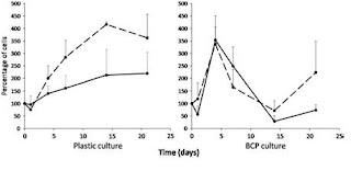
After the first day and some manual homogenization, the authors report seeing a thin monolayer of BCP particle aggregation on the BCP seeded cultures. A scanning electron microscope image was taken of the 3D structures in the cultures after 14 day, revealing a intricate ECM formation that covered the BCP particles; this structure remained throughout the entire experiment. The proliferation assay data shows an increase in hMSC cells up until the 4th day for the BCP cultures in both and osteogenic and proliferative medium, while interestingly the cells proliferate until the 14th day on the PSTC plastic. The authors suggest that this data implies mass osteoblastic differentiation of the hMSCs due to the 3D environment. Moreover, their ALP assay results seemingly affirms this position; the data shows that the cells seeded on BCP particles without osteogenic factors show higher expression of ALP than the 2D plastic cultures. But because ALP expression does not undoubtedly confirm differentiation, so they turn to their RT-PCR data. According to the RT-PCR, in proliferative media, there was higher expression of BMP-2 and BSP on the BCP particles compared to the plastic. And as expected the osteogenic medium cultures showed higher expression of ALP and BSP than the proliferative medium, except for BMP-2. Interestingly, the plastic culture with osteogenic medium data looked very similar to the BCP particle with proliferative medium data.


So the authors finally conclude that the hMSCs can attach to BCP, proliferate, and form 3D structure that ultimately leads to osteoblastic differentiation.

My main complaints are that the authors fail to explain the lower expression of BMP-2 in the osteogenic medium, and they completely neglect the increased proliferation of hMSCs on the BCP particles after day 14 (Figure 2). More nit-picky points are that they choose a very small sample of cells from 3 healthy patients and they only reproduced their experiments 3 times. A suggestion might be to do a quantitative protein assay to look at the existence of protein and their concentration because mRNA does not always mean protein.


Figure 2 hMSC proliferation on different substrates. A fraction of cells was culured on PSTC plastic and another was cultured on BCP ceramic particles in proliferative (dotted lines) or osteogenic medium (full lines) during 21 days.


Figure 3 SEM of 3D environments after 14 day