Monday, March 31, 2008

Bioactive Nanofibers: Synergistic Effects of Nanotopography and Chemical Signaling on Cell Guidance

In this paper, the authors look at the ability of bioactive nanofiber scaffolds to guide cells. The authors specifically investigated the capability of these scaffolds to affect neuronal outgrowth and skin cell migration. Neuronal outgrowth is of particular interest because directed neuronal regeneration has vast medical potential. Skin cell migration is an important component of wound healing, another process of medical interest.
By aligning nanofibers, researchers created a nanotopography that the cells responded to. Compared to random fibers, aligned fibers had much improved neuronal outgrowth and skin cell migration. In addition, by immobilizing specific growth factors on the nanofibers, researchers further increased neuronal outgrowth and skin cell migration.
The experimenters used at ex vivo model to measure neuronal outgrowth, culturing harvested dorsal root ganglia on the scaffold in media. The synergistic effect of nanofiber alignment and growth factor immobilization can be seen in the attached figure from the paper.


On random fibers with no growth factors (A), there was no neurite outgrowth. On aligned, untreated fibers (B), moderate neuronal outgrowth occurred in the direction of alignment. For aligned, bioactive fibers (C), neuronal outgrowth was the greatest. These results demonstrate the importance of both nanotopography and cell signaling for neuronal outgrowth.
An in vitro model was used to measure dermal fibroblast migration. Cells were seeded as monolayers on the nanofiber sheets, and a wound was simulated along the length of the sheet. Researchers then recorded the migration of the dermal fibroblasts into the "wound" site. Cell migration was greatly increased along the axis of alignment. Again, bioactive fibers added to the effect, but not as extensively as in the case of neuronal outgrowth.
This paper is of interest to my group as we will be using nanofibers as a scaffold in our project. We will also be using growth factors, though we will use solubilized growth factors because it is more simple and more effective. This paper uses techniques for visualization that we might find useful for our own project, as we will be using the same scaffolding material, and demonstrates methods for growing cells on nanofiber sheets. In addition, the results of the paper are interesting to me because they offer a viable method of controlling cell migration and neuronal outgrowth, which could be useful in many tissue engineering applications.

Induction of T cell development and establishment of T cell competence from embryonic stem cells differentiated in vitro

Summary:

The authors of the paper tried to direct differentiation of embryonic stem cells into mature and functional T cells by co-culturing them with stromal cells which express Delta-Like 1 (DL1) ligand, initiating the Notch receptor pathways in the cell. In the experiment, embryonic stem cells were grown on two stromal cell lines (OP9-control and OP9-DL1, these cell lines were created by injecting either empty or DL1/GFP containing viral vectors into OP9 cells). Flow cytometry performed on the cells at days 8, 14, and 20 reveal the development and differentiation stages of the embryonic stem cells into lymphocytes. In the presence of DL1 ligand expression, ESCs differentiates into T cells. PCR analysis of these ESC-derived T cells shows that they rearranged during development and have a diverse repertoire of rearranged TCRs (T cell receptors). Upon further investigation, the authors, however, discovered that the majority of these ESC-derived T cells express CD8 only (CD8 identifies T cells as Cytotoxic T Lymphocytes or CTLs, which kill infected cells upon stimulation) and the other major group of T cells (CD4 T cells, which help activate B cells and CTLs), is absent. The experimenters then seeded T cell progenitors into irradiated Fetal Thymic Organ Cultures (FTOCs) and the progenitors were able to reconstitute the FTOC to produce both CD8 and CD4 T cells. These T cells were also shown to mount an antigen-specific response to the LCMV virus.

I am interested in this paper because it shows hope for patients with immunodeficiency, such as HIV, SCID, etc. One reason for HIV’s lethality is the virus’ potent attack against CD4 T cells or T helper cells. CD4 T cells serve one of the most important roles in our immune system including recruiting and stimulate proliferation of B cells and T killer cells. This paper shows that we can use ESCs and direct them to differentiate into functional and effective T cells (including CD4 if transplanted into a FTOC). Once the method is improved in in vitro reconstitution, ESC-derived T cells can be generated in vivo in implanted fetal thymic lobes. By allowing early T cell progenitor to grow in the body, these ESC-derived cells can undergo positive and negative selection. As a result, these cells become self-tolerant and can no longer raise inappropriate immune rejection.

Friday, March 28, 2008

Johnson KR, Leight JL, Weaver VM. Demystifying the effects of a three-dimensional microenvironment in tissue morphogenesis.

In this paper, the researchers mainly focuses on the relationship between the surrounding environment (matrix) and tissue/cell morphogenesis and homeostasis. By understanding how tissues and cells' behavior changes in response to its 3D environment will aid the construction of tissues in vivo that actually would develop and function like the model tissue or organ. In their study, 3D organotypic models are created to study the mechanisms of epithelial morphogenesis and tissue homeostasis. As a result, the scientists found out that there is a critical role being played by the microenvironment in regulating the function and signaling of mammalian tissue.


The experiment designed by the researcher seeks to explore the role of extracellular matrix signaling and tissue organization in epithelial survival. Using breast cancer cells, the experiment shows mammary tissues can resist apoptotic stimuli by activating NF-B through 64 integrin-dependent Rac-Pak1 signaling (occurs in extracellular matrix) and thus, emphasize the importance of the extracellular matrix stroma in tissue survival and suggest that 64 integrin-dependent Rac stimulation of Pak1 could be an important mechanism mediating apoptosis-resistance in some breast tumors.


The experiment make use of two non-malignant human MEC models and a reconstituted, laminin-rich rBM by growing MECs on top of rBM (2D culture) and compared their death-resistance behavior with that of mammary acini embedded within rBM (3D culture). As result, the researchers compared the cells grown as 2D monolayers and epithelial cells assembled into 3D spheroids; finding that 3D assembled cells are more resistant to apoptosis to a degree that is similar to that exhibited by multidrug-, immune- and radiation-resistant tumors. Accordingly, the increased survival behavior of 3D spheroids has been largely attributed to compromised drug penetration and altered cell-cycle dynamics. With this, we can see the importance of the extracellular matrix context in the regulation of homeostasis and growth of cells and tissues.


This paper is very suitable for our project as we also like to compare the matrix and cell growth rate. In our group project, we seek to explore the effect of extracellular matrix stiffness and various anti-cancer drugs to the growth of brain tumor cells. We plan to make different extracellular environments mainly by altering the stiffness of the matrix to which we implant the cells in. Furthermore, the addition of some anti-cancer drugs would demonstrate how easily the drug can get to the cells through the matrix. As a result, we can see the status of the tumor cells via live and dead assay to draw conclusion about the growth rate/destruction of the brain tumor cells.

Thursday, March 27, 2008

Tissue Engineering of Vascularized Cardiac Muscle From Human Embryonic Stem Cells

Abstract:
Transplantation of a tissue-engineered heart muscle represents a novel experimental therapeutic paradigm for myocardial diseases. However, this strategy has been hampered by the lack of sources for human cardiomyocytes and by the scarce vasculature in the ischemic area limiting the engraftment and survival of the transplanted muscle. Beyond the necessity of endothelial capillaries for the delivery of oxygen and nutrients to the grafted muscle tissue, interactions between endothelial and cardiomyocyte cells may also play a key role in promoting cell survival and proliferation. In the present study, we describe the formation of synchronously contracting engineered human cardiac tissue derived from human embryonic stem cells containing endothelial vessel networks. The 3D muscle consisted of cardiomyocytes, endothelial cells (ECs), and embryonic fibroblasts (EmFs). The formed vessels were further stabilized by the presence of mural cells originating from the EmFs. The presence of EmFs decreased EC death and increased EC proliferation. Moreover, the presence of endothelial capillaries augmented cardiomyocyte proliferation and did not hamper cardiomyocyte orientation and alignment. Immunostaining, ultrastructural analysis (using transmission electron microscopy), RT-PCR, pharmacological, and confocal laser calcium imaging studies demonstrated the presence of
cardiac-specific molecular, ultrastructural, and functional properties of the generated tissue constructs with synchronous activity mediated by action potential propagation through gap junctions. In summary, this is the first report of the construction of 3D vascularized human cardiac tissue that may have unique applications for studies of cardiac development, function, and tissue replacement therapy.

Response:
It is without a doubt that there is need for artificial tissue and organs to battle against a myriad of human problems and complications with the body. It was amazing to me how this engineered heart tissue (EHT) was shown to beat and pulse synchronously and show similarities to that of real human hearts. This particular article interested me because they were able to create the first 3D, vascularized human cardiac tissue, which has multiple implications-- using it as a model for studying the human heart in vitro, as well as a huge advancement in tissue replacement therapy. This vascularization is important because all previous EHTs showed poor survival of seeded cells (myocytes) in the scaffold. Caspi then hypothesized that seeding endothelial cells, as well as embryonic fibroblasts, will improve vascularization in the EHT and improve cardiomyocyte growth and viability. Immunohistochemistry was used to show that the tri-cultured scaffolds had a higher density of vascularization, and immunoflourescent staining was used to show that the vascular networks in these scaffolds showed structural organization. RT-PCR was used to show that the cardiomyocytes in the tri-cultured scaffold were mature and differentiated-- more so than the controls. Other assays were used to analyze cell growth and viability, but you can read the paper for that. So, I thought this was a pretty thorough and well-done experiment with very promising potentials in the medical and research world. The only problem I had with it was that they don't go into how the addition of EC's and EmF's promote vascularization and survival--that is, the interactions, signalling pathways, etc. that allow for this to happen, but it is mentioned that this particular topic is currently being researched and offers potential answers.

Wednesday, March 26, 2008

Tissue Engineering of Complex Tooth Structures on Biodegradable Polymer Scaffolds


C.S. Young1, S. Terada2, J.P. Vacanti2, M. Honda3, J.D. Bartlett1*, and P.C. Yelick1* 1Department of Cytokine Biology and Harvard-Forsyth Department of Oral Biology, The Forsyth Institute, Boston, MA 02115, USA; 2Department of Surgery, Massachusetts General Hospital and Harvard Medical School, Boston, MA, USA; and 3Department of Oral and Maxillofacial Surgery, Nagoya University School of Medicine, Nagoya, Japan; *corresponding authors, pyelick@forsyth.org, jbartlett@forsyth.org | J Dent Res 81(10):695-700, 2002 (http://jdr.iadrjournals.org/cgi/content/abstract/81/10/695)

Wh
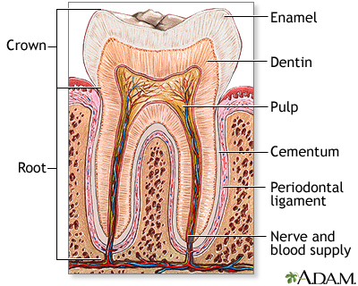
y Study Tooth Tissue Engineering? (other than the fact that teeth are simply awesome!)

Trauma, dental caries, and a variety of genetic disorder such as amelogenesis imperfecta can result in a tooth loss, for which there is no regenerative biological substitute currently. This particular area of research has been flourishing since the Forsyth Institute (Boston, MA) presented a possibility of generating bioengineered dental tissues after they found that bioengineered dental tissues were constructed using mixed population of cultured post-natal tooth bud cells (British Dental Journal (2006); 201, 73. doi: 10.1038/sj.bdj.4813885). In order to investigate a tissue engineering approach to construct a biologically suitable substitute for a tooth, this paper I chose (coincidentally it’s from the Forsyth Institute) examines a tissue engineering approach that was used to bioengineer small intestines successfully (Choi and Vacanti, 1997).

What does this study aim to discover in order to demonstrate a regenerative tooth crown?

The signaling between dental mesenchyme that differentiates into pulp and dentin tissues and epithelial tissue of the enamel that produces the dental enamel plays a pivotal role in the overall development of teeth: allowing each tooth to possess different characteristics such as its specific size, shape and function within different positions in the jaw. Thus, this study aims to find the presence of epithelial and mesenchymal dental stem cells in porcine third molar tissues that allow for a successful construction of tooth crowns from dissociated tooth tissues.

*Summary*

Biodegradable polymer scaffolds (a human tooth-shaped polymer scaffolds made with PGA/PLLA and PLGA) were used as substrates to seed dissociated cells from tooth tissues. Third molar tooth buds were obtained from 6 months old pig jaws; pulp organ tissues and enamel tissues were minced after. This cell/polymer constructs were implanted into a suitable host (rat) to allow for the growth of higher-ordered structures. Histological, immunohistochemical, and Laser-capture Microdissection and Reverse-transcription/Polymerase Chain-reaction (LCM, RT-PCR) analyses were on samples from 20, 25, and 30-weeks of implants.

Overall, molecular evidences found from the noted analytical methods indicated a successful bioengineering of complex tooth crowns that closely resemble naturally developing teeth. After 20-weeks of implanting, mineralized dentin, pre-dentin, and pulp tissue resembling vascularized mesenchyme were present. Amelogenin, one of the most prominent extracellular matrix proteins in developing enamel was immunodetected in the enamel matrix of 25-week implant as well as the presence of ameloblast-like cells adjacent to the enamel. 30-week implant tissues showed a thick layer of enamel surrounding a layer of dentin. Amelogenin was detected at the secretory ends of ameloblast-like cells, confirming the occurrence of amelogenesis in the developing enamel. LCM and RT-PCR using nested primers for odontoblast-specific DSPP mRNA confirmed the identity of putative odontoblast-like cells present in bioengineered tooth tissues. Given these morphological, histological, and immunochemical evidences from 20-, 25-, and 30-week implant tissues, this study overall demonstrated a successful construction of bioengineered teeth containing the similar organization and molecular markers of natural teeth such as dentin, pulp, and amelogenin proteins.

Why Did I Choose this Paper?

The approach (seeding dissociated tooth tissues onto biodegradable scaffolds) this paper took signifies a possibility of generating teeth of preprogrammed size and shape, giving engineers more control over the overall function and morphology of bioengineered tooth. The bioengineered tissues not only provided morphologically evidences of natural teeth, LCM and RT-PCR further confirmed the on-going process of amelogenesis. Since my research interest lies within dental science, specifically in understanding amelogensis by closely mimicking the enamel formation in vitro, I chose this paper to study possible approaches engineers can take in regenerating teeth that closely resemble the natural teeth. If we can understand how biological system is able to design organized structures (ie. enamel) in tissues at the molecular level, not only people with genetically impaired teeth could recover their full dentition but we may even be able to use artificial human enamel to build a dental restoration. For instance, if the molecular mechanism of amelogensis was known, engineers can further manipulate the developmental process of bioengineered teeth to produce fully-developed enamel.

*Here are some interesting papers you can read at your leisure:

  • Micelle Structure of Amelogenin in Porcine Secretory Enamel: J Dent Res 86(8):758-763, 2007 (http://jdr.iadrjournals.org/cgi/content/abstract/86/8/758)
  • Bioengineered Teeth from Cultured Rat Tooth Bud Cells: J Dent Res 83(7): 523-528, 2004 (http://jdr.iadrjournals.org/cgi/content/full/83/7/523)
  • New observations of the hierarchical structure of human enamel, from nanoscale to microscale: Journal of Tissue Engineering and Regenerative Medicine V1 I3: 185-191 (http://www3.interscience.wiley.com/cgi-bin/abstract/114219401/ABSTRACT)

Friday, March 21, 2008

Tissue Engineering of the Meniscus

The meniscus is crucial in bearing and distributing loads, absorbing shock, lubricating, and stabilizing the knee. Injuries to the meniscus are some of the most common injuries seen in orthopaedics and often lead to osteoarthritis and degradation of articular cartilage. More promising means to repair meniscal tears or synthesize an artificial meniscus are necessary, because the meniscus has a limited regenerative capability and injuries often occur in the interior, avascular region of the meniscus. Tissue engineering offers new treatment possibilities for meniscal repairs and replacements.

The phenotype of the meniscal tissue is similar to fibro-cartilage in the avascular region and is fibrous in the vascular region, consisting predominantly of type I collagen and glycosaminoglycans. Scaffolds that are biocompatible and biodegradable, allow unrestricted cell growth and free diffusion of nutrients, can be used as a carrier for growth factors, and maintain structural integrity while supporting the joint on the knee are qualifications for an ideal scaffold material. Scaffolds based on collagen molecules are the most promising, because of their optimal load-bearing capacity and the control of pore creation for new tissue ingrowth. Using whole tissues or isolated tissue components as scaffold material are often disadvantageous, because they cannot often bear large loads. The use of synthetic polymer-based scaffolds is currently being investigated because of the ability to control the porosity, the degradation rate, and other mechanical properties of polymers. In polymer design, the adhesive potential of the polymer scaffold to the host tissue is of key importance.

Other potential cells for investigation in scaffold associated studies involve nondifferentiated progenitor cells or fibroblasts, which have differentiated into fibro-cartilage-like cells in previous studies. Additionally, growth factors which stimulate synthesis and inhibit the degradation of the ECM have been studied for their effects on cultured meniscal cells or meniscus explants. Such growth factors include TGF-β, PDGF, IGF-I, which stimulate glycosaminoglycan production, proliferation of meniscal cells, and production of cartilage matrix molecules, respectively. As for replacing a meniscus with an autologous allograft, problems occur because of poor initial mechanical properties of the materials, preventing the allograft from lasting long-term. Donor menisci implants have associated problems, such as immunological reactions, disease transfer, and reshaping of the implant to fit the patient. More importantly, such implants often induce high friction in the knee joint, resulting in damage to the cartilage.

Meniscal injuries are exceedingly common in athletes, particularly soccer players and runners. If the meniscus degenerates or is removed, the remaining cartilage in the knee joint slowly wears away, leading to increasing pain in the joint, osteoarthritis, and severe joint degeneration. At this point, artificial joint replacement seems to be the most promising way to relieve the pain of osteoarthritis. However, there are numerous problems with this painful procedure, especially the length of time the implant lasts, usually around ten years, and the stresses on the bone that the implant places. Repairing the meniscus or offering new treatments using synthetic tissue engineering, provides these athletes with a remedy for their knee problems, enabling them to live with less pain, avoid artificial joint replacements, and possibly return to their sport. Currently recovering from my second meniscal tear, I am especially interested in new technologies and research into synthetic menisci and more effective treatments for meniscal repairs.

Wednesday, March 19, 2008

Enhanced differentiation of mesenchymal stem cells co-cultured with ligament fibroblasts on gelatin/silk fibroin hybrid scaffold

Enhanced differentiation of mesenchymal stem cells co-cultured with ligament fibroblasts on gelatin/silk fibroin hybrid scaffold

H. Fan, H. Liu, S.L. Toh, and J.C.H. Goh, Biomaterials 29 (2008), pp. 1017-1027.

In this paper, MSCs were co-cultured with anterior cruciate ligament (ACL) fibroblasts to determine whether soluble signals released by the fibroblasts enhanced the differentiation of the MSCs into fibroblasts. The motivation behind this research is to be able to use such a co-culture system to create a tissue-engineered ligament in vitro to remedy ACL injuries.

MSCs were harvested from rabbit bone marrow and seeded onto silk cable-reinforced gelatin/silk fibroin scaffolds. After incubation, the constructs were transferred into co-culture systems with fibroblasts (isolated from the ACL of the same rabbits) or into non-co-culture systems (control). Over a two-week culture period, observations for the following were made:
1) Cell proliferation (by quantifying DNA content with Hoechst Dye and a fluorescent plate reader)
2) Cell metabolism (by alamar blue assay)
3) Cell viability (by FDA/PI staining)
4) Cell morphology (by SEM)
5) Collagen production (by Sircol collagen dye binding assay)
6) Gene expression of collagen I, collagen III, and tenascin-C (by quantitative RT-PCR)
In addition, histological slides were prepared as well as a Western blot for collagen I, collagen III, and tenascin-C.

It was found that the MSCs in the co-cultures developed fibroblast-like morphology. Also, the mRNA and protein levels of collagen I, collagen III, and tenascin-C were significantly higher for the MSCs in the co-cultures than for the non-co-cultures. These results support the hypothesis that the 3-D co-culture of MSCs with ACL fibroblasts facilitated the differentiation of the MSCs.

I chose this paper because it discusses co-culturing as an alternative to the usual biochemical and mechanical stimulation of MSC differentiation. Co-culturing is certainly more efficient and cost-effective than having to figure out and to obtain the proper growth factors to induce differentiation.

Overall, I liked this paper because the authors fairly thoroughly characterized the behavior of the MSCs. Their protocol was clearly detailed and easy to understand (especially since we've already discussed each of these techniques in this class). However, I do wish that they had gone one step further and characterized some of the mechanical properties of their tissue-engineered constructs.

Monday, March 17, 2008

Cell Shape, Cytoskeletal Tension, and RhoA Regulate Stem Cell Lineage Commitment

Cell Shape, Cytoskeletal Tension, and RhoA Regulate Stem Cell Lineage Commitment
Rowena McBeath, Dana M. Pirone, Celeste M. Nelson, Kiran Bhadriraju, and Christopher S. Chen.
Developmental Cell, Vol. 6, 483–495, April, 2004.

Summary:
Commitment of stem cells to different lineages is regulated by many cues in the local tissue microenvironment. Here we demonstrate that cell shape regulates commitment of human mesenchymal stem cells (hMSCs) to adipocyte or osteoblast fate. hMSCs allowed to adhere, flatten, and spread underwent osteogenesis, while unspread, round cells became adipocytes. Cell shape regulated the switch in lineage commitment by modulating endogenous RhoA activity. Expressing dominant-negative RhoA committed hMSCs to become adipocytes, while constitutively active RhoA caused osteogenesis. However, the RhoA-mediated adipogenesis or osteogenesis was conditional on a round or spread shape, respectively, while constitutive activation of the RhoA effector, ROCK, induced osteogenesis independent of cell shape. This RhoA- ROCK commitment signal required actin-myosin-generated tension. These studies demonstrate that mechanical cues experienced in developmental and adult contexts, embodied by cell shape, cytoskeletal tension, and RhoA signaling, are integrato the commitment of stem cell fate.


I find this paper really interesting because it demonstrates how physical cues like island size affects cell shape, which in turn affects cell's ability to make decisions about its fate. The authors showed that cell shape acts as a mechanical cue in driving hMSC commitment to osteoblasts vs. adipocytes. I also liked how the authors were able to study the transduction of these physical signals into biochemical processes by doing constitutively active/inactive experiments. Additionally, the authors use techniques such as micro-island patterning, FACS, RT-PCR, recombinant adenovirus constructs etc. to study this bioengineering problem.

Here is the link for google scholar:
http://scholar.google.com/scholar?q=http%3A%2F%2Flinkinghub.elsevier.com%2Fretrieve%2Fpii%2FS1534580704000759&hl=en&lr=&btnG=Search

Sunday, March 16, 2008

Evoked Acetylcholine Release by Immortalized Brain Endothelial Cells Genetically Modified...

Evoked Acetylcholine Release by Immortalized Brain Endothelial Cells Genetically Modified to Express Choline Acetyltransferase and/or the Vesicular Acetylcholine Transporter

M. Malo, M.-F. Diebler, L. Prado de Carvalho, F.-M. Meunier, *Y. Dunant, *A. Bloc, J. Stinnakre, M. Tomasi, †J. Tche´linge´rian, †P. O. Couraud, and M. Israe¨l

The goal of the study was to genetically modify RBE4 cells, rat brain endothelial cells, so that not only do they produce ACh but package it into an internal store, as to mimic the vesicular store if neurotransmitters in neurons. RBE4 cells can’t accumulate ACh but can release it upon calcium entry. RBE4 were used because they already normally have machinery specific to the release of ACh (and no other neurotransmitter) and they are convenient for gene manipulations.

In this experiment, two things were done:
(1) The RBE4 cells were engineered to express ChAT, choline acetyltransferase. (Those RBE4 cells that expressed ChAT produced endogenous Ach).
(2) The experimenters attempted to modify the RBE4 cells expressing ChAT further to express the vesicular Ach transporter (VAChT).

Gene modification was done with the RBE4 cells in suspension with a transfection reagent, a selection vector, a ChAT expression vector and a VAChT expression vector. ChAT activity was determined by radiolabeling the ACh and was quantified using the Bradford assay. VAChT expression was assessed via a binding assay with vesamicol as specific ligand. An immunodetection of ChAT and VAChT was done and visualized with a chemiluminescent system. Results of these tests showed that stable transfected RBE4 cells only expressed either ChAT or VAChT, not both (but transfection overall was high). Thus, the effects of VAChT on ACh release could only be tested through evoked ACh release after passive loading of the VAChT-expressing cells with ACh in comparison to the ChAT-expressing cells and no significant difference was seen.

A chemiluminescent procedure was also used to quantify ACh release. Electrophysiologically, ACh release was measured by measuring the membrane current of Xenopus myocytes (used as detector cells). It was seen that with calcium addition, ACh was released. Synaptic-like currents were also observed. Electrophysiological testing of VAChT expressing cells suggested that temporal pattern of ACh release was controlled by VAChT. Why this is was not answered in this experiment.

I chose this paper because I'm particularly interested in neurons and the experiment includes techniques that we have learned in class. It is important because the researchers were able to induce synthesis and accumulation of ACh in non-neuronal cells. This may be a great treatment for neurodegenerative diseases that lead to cholinergic deficits.

Wednesday, March 12, 2008

Promotion of Osteogenesis in Tissue-Engineered Bone by Pre-Seeding Endothelial Progenitor Cells-Derived Endothelial Cells

This paper investigates the potential benefits of seeding endothelial cells onto tissue-engineered bone. Given a biocompatible scaffold and an osteogenic cell population, a vascular bed would help to overcome the difficulties of nutrient and oxygen transport in the 3D implant. The investigators tested their hypothesis that EC's help promote vascularization by using three test groups. First, osteoblasts and EC's were differentiated from the bone arrow of BALB/c mice. They used a porous polycaprolactone (PCL)-hydroxyapatite(HA) scaffold as the test implant.  One test group was the control (cell-free), one group was implants with only osteoblasts and the last group was implants with osteoblasts and EC's seeded onto it. The implants were placed in a 0.4cm long segmental femur defect. 

Evaluating the grafts histologically, the investigators found that after 6 weeks of implantation, the EC-OB group had a much more widely distributed capillary network. It also had osteoid generated by osteoblasts and ischemic necroses was absent. The conclusion of the paper is that pre-seeding implants with EC's did effectively promote neovascularization in grafts and improved osteogenesis while avoiding necrosis.
I believe this paper is important because it investigates methods of making tissue-engineered bone implants more effective in vivo. Now that scientists have found ways of making structurally sound scaffolds that are compatible with the body, the real challenge is to make this implant bioactive rather than just inert. This a key component to making tissue-engineered bone successful.