Wednesday, April 02, 2008

Neural Tissue Engineering: a Self Organizing Collagen Guidance Conduit

This paper discusses a new method of creating 3-D neural tissue using seeded collagen gels for self-alignment of the tissue. Within the medical community, nerve autografts are the "gold standard," but with the development of this nerve tissue method there is a potential for surgical implantation without removal of other nerve cells that damage donating nerves. Previous studies have shown that collagen gels are an effective way of attaining 3-D tissue cultures, therefore the researchers are applying this knowledge to create nerve cultures that have aligned schwann cells to match true nerve abilities both physically and biologically.


This research used collagen gels (rectangular)seeded with 25,000 schwann cells (prepared from male Fischer rats) per gel. Then dorsal root ganglia (taken from Sprague adult rats') were prepared using DMEM with collagenase. They were later applied to the collagen gels for adherence and growth.

After a 3 day incubation, the gels were washed, and stained with paraformaldehyde to detect betaIII-tubulin, a marker for neurons. Antibodies followed by secondary antibody allowed observation with fluorescence microscope.


The contruction of a device to mimic a nerve began with a silicone tube with eight holes for the collagen seeded gels to be pushed through (like a push pop). This device was inserted into female rat precisely damaged sciatic nerve for in vivo growth studies. If the inserted collagen moved away from the walls of the silicone tube, then self alignment would be considered successful.


The quantification of realignment and growth were performed using transverse and longitudinal sections taken from the insertion site along with the use of fluorescence microscopy.


The results reported showed that the schwann cells within the collagen gell had become aligned with the axis of the tube and DRG neurons extended paralled to the tube walls.


Evaluation of regeneration of a transverse section using the "collagenated tube", no inserted device and empty silicone tube showed that the collagenated had the greatest repairing abilities over time with the most growth.

Longitudinal sections showed the same results with most observed regeneration in the collagenated tubes.
Factors that may influenced results may include variation between rat species, age and sex. The age of the source of DRG may have influeced the potential for regrowth. The change from male to female for the in vivo test may also have affected the amount of regeneration and the direction of the alignment.
This paper presents a good protocal for the development of neural tissue to have correct alignment, essential for nerve communication. This paper was interesting especially after reading previous posts about tissue regeneration. Protocols for tissue development used collagen gels and this further exemplified the potential for 3-D culture. This paper caught my attention because of its attention to detail of the protocol and the future application of this device for treatment of patients with nerve damage such as from car accidents or war injuries.

Monday, March 31, 2008

Bioactive Nanofibers: Synergistic Effects of Nanotopography and Chemical Signaling on Cell Guidance

In this paper, the authors look at the ability of bioactive nanofiber scaffolds to guide cells. The authors specifically investigated the capability of these scaffolds to affect neuronal outgrowth and skin cell migration. Neuronal outgrowth is of particular interest because directed neuronal regeneration has vast medical potential. Skin cell migration is an important component of wound healing, another process of medical interest.
By aligning nanofibers, researchers created a nanotopography that the cells responded to. Compared to random fibers, aligned fibers had much improved neuronal outgrowth and skin cell migration. In addition, by immobilizing specific growth factors on the nanofibers, researchers further increased neuronal outgrowth and skin cell migration.
The experimenters used at ex vivo model to measure neuronal outgrowth, culturing harvested dorsal root ganglia on the scaffold in media. The synergistic effect of nanofiber alignment and growth factor immobilization can be seen in the attached figure from the paper.


On random fibers with no growth factors (A), there was no neurite outgrowth. On aligned, untreated fibers (B), moderate neuronal outgrowth occurred in the direction of alignment. For aligned, bioactive fibers (C), neuronal outgrowth was the greatest. These results demonstrate the importance of both nanotopography and cell signaling for neuronal outgrowth.
An in vitro model was used to measure dermal fibroblast migration. Cells were seeded as monolayers on the nanofiber sheets, and a wound was simulated along the length of the sheet. Researchers then recorded the migration of the dermal fibroblasts into the "wound" site. Cell migration was greatly increased along the axis of alignment. Again, bioactive fibers added to the effect, but not as extensively as in the case of neuronal outgrowth.
This paper is of interest to my group as we will be using nanofibers as a scaffold in our project. We will also be using growth factors, though we will use solubilized growth factors because it is more simple and more effective. This paper uses techniques for visualization that we might find useful for our own project, as we will be using the same scaffolding material, and demonstrates methods for growing cells on nanofiber sheets. In addition, the results of the paper are interesting to me because they offer a viable method of controlling cell migration and neuronal outgrowth, which could be useful in many tissue engineering applications.

Induction of T cell development and establishment of T cell competence from embryonic stem cells differentiated in vitro

Summary:

The authors of the paper tried to direct differentiation of embryonic stem cells into mature and functional T cells by co-culturing them with stromal cells which express Delta-Like 1 (DL1) ligand, initiating the Notch receptor pathways in the cell. In the experiment, embryonic stem cells were grown on two stromal cell lines (OP9-control and OP9-DL1, these cell lines were created by injecting either empty or DL1/GFP containing viral vectors into OP9 cells). Flow cytometry performed on the cells at days 8, 14, and 20 reveal the development and differentiation stages of the embryonic stem cells into lymphocytes. In the presence of DL1 ligand expression, ESCs differentiates into T cells. PCR analysis of these ESC-derived T cells shows that they rearranged during development and have a diverse repertoire of rearranged TCRs (T cell receptors). Upon further investigation, the authors, however, discovered that the majority of these ESC-derived T cells express CD8 only (CD8 identifies T cells as Cytotoxic T Lymphocytes or CTLs, which kill infected cells upon stimulation) and the other major group of T cells (CD4 T cells, which help activate B cells and CTLs), is absent. The experimenters then seeded T cell progenitors into irradiated Fetal Thymic Organ Cultures (FTOCs) and the progenitors were able to reconstitute the FTOC to produce both CD8 and CD4 T cells. These T cells were also shown to mount an antigen-specific response to the LCMV virus.

I am interested in this paper because it shows hope for patients with immunodeficiency, such as HIV, SCID, etc. One reason for HIV’s lethality is the virus’ potent attack against CD4 T cells or T helper cells. CD4 T cells serve one of the most important roles in our immune system including recruiting and stimulate proliferation of B cells and T killer cells. This paper shows that we can use ESCs and direct them to differentiate into functional and effective T cells (including CD4 if transplanted into a FTOC). Once the method is improved in in vitro reconstitution, ESC-derived T cells can be generated in vivo in implanted fetal thymic lobes. By allowing early T cell progenitor to grow in the body, these ESC-derived cells can undergo positive and negative selection. As a result, these cells become self-tolerant and can no longer raise inappropriate immune rejection.

Friday, March 28, 2008

Johnson KR, Leight JL, Weaver VM. Demystifying the effects of a three-dimensional microenvironment in tissue morphogenesis.

In this paper, the researchers mainly focuses on the relationship between the surrounding environment (matrix) and tissue/cell morphogenesis and homeostasis. By understanding how tissues and cells' behavior changes in response to its 3D environment will aid the construction of tissues in vivo that actually would develop and function like the model tissue or organ. In their study, 3D organotypic models are created to study the mechanisms of epithelial morphogenesis and tissue homeostasis. As a result, the scientists found out that there is a critical role being played by the microenvironment in regulating the function and signaling of mammalian tissue.


The experiment designed by the researcher seeks to explore the role of extracellular matrix signaling and tissue organization in epithelial survival. Using breast cancer cells, the experiment shows mammary tissues can resist apoptotic stimuli by activating NF-B through 64 integrin-dependent Rac-Pak1 signaling (occurs in extracellular matrix) and thus, emphasize the importance of the extracellular matrix stroma in tissue survival and suggest that 64 integrin-dependent Rac stimulation of Pak1 could be an important mechanism mediating apoptosis-resistance in some breast tumors.


The experiment make use of two non-malignant human MEC models and a reconstituted, laminin-rich rBM by growing MECs on top of rBM (2D culture) and compared their death-resistance behavior with that of mammary acini embedded within rBM (3D culture). As result, the researchers compared the cells grown as 2D monolayers and epithelial cells assembled into 3D spheroids; finding that 3D assembled cells are more resistant to apoptosis to a degree that is similar to that exhibited by multidrug-, immune- and radiation-resistant tumors. Accordingly, the increased survival behavior of 3D spheroids has been largely attributed to compromised drug penetration and altered cell-cycle dynamics. With this, we can see the importance of the extracellular matrix context in the regulation of homeostasis and growth of cells and tissues.


This paper is very suitable for our project as we also like to compare the matrix and cell growth rate. In our group project, we seek to explore the effect of extracellular matrix stiffness and various anti-cancer drugs to the growth of brain tumor cells. We plan to make different extracellular environments mainly by altering the stiffness of the matrix to which we implant the cells in. Furthermore, the addition of some anti-cancer drugs would demonstrate how easily the drug can get to the cells through the matrix. As a result, we can see the status of the tumor cells via live and dead assay to draw conclusion about the growth rate/destruction of the brain tumor cells.

Thursday, March 27, 2008

Tissue Engineering of Vascularized Cardiac Muscle From Human Embryonic Stem Cells

Abstract:
Transplantation of a tissue-engineered heart muscle represents a novel experimental therapeutic paradigm for myocardial diseases. However, this strategy has been hampered by the lack of sources for human cardiomyocytes and by the scarce vasculature in the ischemic area limiting the engraftment and survival of the transplanted muscle. Beyond the necessity of endothelial capillaries for the delivery of oxygen and nutrients to the grafted muscle tissue, interactions between endothelial and cardiomyocyte cells may also play a key role in promoting cell survival and proliferation. In the present study, we describe the formation of synchronously contracting engineered human cardiac tissue derived from human embryonic stem cells containing endothelial vessel networks. The 3D muscle consisted of cardiomyocytes, endothelial cells (ECs), and embryonic fibroblasts (EmFs). The formed vessels were further stabilized by the presence of mural cells originating from the EmFs. The presence of EmFs decreased EC death and increased EC proliferation. Moreover, the presence of endothelial capillaries augmented cardiomyocyte proliferation and did not hamper cardiomyocyte orientation and alignment. Immunostaining, ultrastructural analysis (using transmission electron microscopy), RT-PCR, pharmacological, and confocal laser calcium imaging studies demonstrated the presence of
cardiac-specific molecular, ultrastructural, and functional properties of the generated tissue constructs with synchronous activity mediated by action potential propagation through gap junctions. In summary, this is the first report of the construction of 3D vascularized human cardiac tissue that may have unique applications for studies of cardiac development, function, and tissue replacement therapy.

Response:
It is without a doubt that there is need for artificial tissue and organs to battle against a myriad of human problems and complications with the body. It was amazing to me how this engineered heart tissue (EHT) was shown to beat and pulse synchronously and show similarities to that of real human hearts. This particular article interested me because they were able to create the first 3D, vascularized human cardiac tissue, which has multiple implications-- using it as a model for studying the human heart in vitro, as well as a huge advancement in tissue replacement therapy. This vascularization is important because all previous EHTs showed poor survival of seeded cells (myocytes) in the scaffold. Caspi then hypothesized that seeding endothelial cells, as well as embryonic fibroblasts, will improve vascularization in the EHT and improve cardiomyocyte growth and viability. Immunohistochemistry was used to show that the tri-cultured scaffolds had a higher density of vascularization, and immunoflourescent staining was used to show that the vascular networks in these scaffolds showed structural organization. RT-PCR was used to show that the cardiomyocytes in the tri-cultured scaffold were mature and differentiated-- more so than the controls. Other assays were used to analyze cell growth and viability, but you can read the paper for that. So, I thought this was a pretty thorough and well-done experiment with very promising potentials in the medical and research world. The only problem I had with it was that they don't go into how the addition of EC's and EmF's promote vascularization and survival--that is, the interactions, signalling pathways, etc. that allow for this to happen, but it is mentioned that this particular topic is currently being researched and offers potential answers.

Wednesday, March 26, 2008

Tissue Engineering of Complex Tooth Structures on Biodegradable Polymer Scaffolds


C.S. Young1, S. Terada2, J.P. Vacanti2, M. Honda3, J.D. Bartlett1*, and P.C. Yelick1* 1Department of Cytokine Biology and Harvard-Forsyth Department of Oral Biology, The Forsyth Institute, Boston, MA 02115, USA; 2Department of Surgery, Massachusetts General Hospital and Harvard Medical School, Boston, MA, USA; and 3Department of Oral and Maxillofacial Surgery, Nagoya University School of Medicine, Nagoya, Japan; *corresponding authors, pyelick@forsyth.org, jbartlett@forsyth.org | J Dent Res 81(10):695-700, 2002 (http://jdr.iadrjournals.org/cgi/content/abstract/81/10/695)

Why Study Tooth Tissue Engineering? (other than the fact that teeth are simply awesome!)

Trauma, dental caries, and a variety of genetic disorder such as amelogenesis imperfecta can result in a tooth loss, for which there is no regenerative biological substitute currently. This particular area of research has been flourishing since the Forsyth Institute (Boston, MA) presented a possibility of generating bioengineered dental tissues after they found that bioengineered dental tissues were constructed using mixed population of cultured post-natal tooth bud cells (British Dental Journal (2006); 201, 73. doi: 10.1038/sj.bdj.4813885). In order to investigate a tissue engineering approach to construct a biologically suitable substitute for a tooth, this paper I chose (coincidentally it’s from the Forsyth Institute) examines a tissue engineering approach that was used to bioengineer small intestines successfully (Choi and Vacanti, 1997).

What does this study aim to discover in order to demonstrate a regenerative tooth crown?

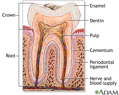
The signaling between dental mesenchyme that differentiates into pulp and dentin tissues and epithelial tissue of the enamel that produces the dental enamel plays a pivotal role in the overall development of teeth: allowing each tooth to possess different characteristics such as its specific size, shape and function within different positions in the jaw. Thus, this study aims to find the presence of epithelial and mesenchymal dental stem cells in porcine third molar tissues that allow for a successful construction of tooth crowns from dissociated tooth tissues.

*Summary*

Biodegradable polymer scaffolds (a human tooth-shaped polymer scaffolds made with PGA/PLLA and PLGA) were used as substrates to seed dissociated cells from tooth tissues. Third molar tooth buds were obtained from 6 months old pig jaws; pulp organ tissues and enamel tissues were minced after. This cell/polymer constructs were implanted into a suitable host (rat) to allow for the growth of higher-ordered structures. Histological, immunohistochemical, and Laser-capture Microdissection and Reverse-transcription/Polymerase Chain-reaction (LCM, RT-PCR) analyses were on samples from 20, 25, and 30-weeks of implants.

Overall, molecular evidences found from the noted analytical methods indicated a successful bioengineering of complex tooth crowns that closely resemble naturally developing teeth. After 20-weeks of implanting, mineralized dentin, pre-dentin, and pulp tissue resembling vascularized mesenchyme were present. Amelogenin, one of the most prominent extracellular matrix proteins in developing enamel was immunodetected in the enamel matrix of 25-week implant as well as the presence of ameloblast-like cells adjacent to the enamel. 30-week implant tissues showed a thick layer of enamel surrounding a layer of dentin. Amelogenin was detected at the secretory ends of ameloblast-like cells, confirming the occurrence of amelogenesis in the developing enamel. LCM and RT-PCR using nested primers for odontoblast-specific DSPP mRNA confirmed the identity of putative odontoblast-like cells present in bioengineered tooth tissues. Given these morphological, histological, and immunochemical evidences from 20-, 25-, and 30-week implant tissues, this study overall demonstrated a successful construction of bioengineered teeth containing the similar organization and molecular markers of natural teeth such as dentin, pulp, and amelogenin proteins.

Why Did I Choose this Paper?

The approach (seeding dissociated tooth tissues onto biodegradable scaffolds) this paper took signifies a possibility of generating teeth of preprogrammed size and shape, giving engineers more control over the overall function and morphology of bioengineered tooth. The bioengineered tissues not only provided morphologically evidences of natural teeth, LCM and RT-PCR further confirmed the on-going process of amelogenesis. Since my research interest lies within dental science, specifically in understanding amelogensis by closely mimicking the enamel formation in vitro, I chose this paper to study possible approaches engineers can take in regenerating teeth that closely resemble the natural teeth. If we can understand how biological system is able to design organized structures (ie. enamel) in tissues at the molecular level, not only people with genetically impaired teeth could recover their full dentition but we may even be able to use artificial human enamel to build a dental restoration. For instance, if the molecular mechanism of amelogensis was known, engineers can further manipulate the developmental process of bioengineered teeth to produce fully-developed enamel.

*Here are some interesting papers you can read at your leisure:

  • Micelle Structure of Amelogenin in Porcine Secretory Enamel: J Dent Res 86(8):758-763, 2007 (http://jdr.iadrjournals.org/cgi/content/abstract/86/8/758)
  • Bioengineered Teeth from Cultured Rat Tooth Bud Cells: J Dent Res 83(7): 523-528, 2004 (http://jdr.iadrjournals.org/cgi/content/full/83/7/523)
  • New observations of the hierarchical structure of human enamel, from nanoscale to microscale: Journal of Tissue Engineering and Regenerative Medicine V1 I3: 185-191 (http://www3.interscience.wiley.com/cgi-bin/abstract/114219401/ABSTRACT)

Friday, March 21, 2008

Tissue Engineering of the Meniscus

The meniscus is crucial in bearing and distributing loads, absorbing shock, lubricating, and stabilizing the knee. Injuries to the meniscus are some of the most common injuries seen in orthopaedics and often lead to osteoarthritis and degradation of articular cartilage. More promising means to repair meniscal tears or synthesize an artificial meniscus are necessary, because the meniscus has a limited regenerative capability and injuries often occur in the interior, avascular region of the meniscus. Tissue engineering offers new treatment possibilities for meniscal repairs and replacements.

The phenotype of the meniscal tissue is similar to fibro-cartilage in the avascular region and is fibrous in the vascular region, consisting predominantly of type I collagen and glycosaminoglycans. Scaffolds that are biocompatible and biodegradable, allow unrestricted cell growth and free diffusion of nutrients, can be used as a carrier for growth factors, and maintain structural integrity while supporting the joint on the knee are qualifications for an ideal scaffold material. Scaffolds based on collagen molecules are the most promising, because of their optimal load-bearing capacity and the control of pore creation for new tissue ingrowth. Using whole tissues or isolated tissue components as scaffold material are often disadvantageous, because they cannot often bear large loads. The use of synthetic polymer-based scaffolds is currently being investigated because of the ability to control the porosity, the degradation rate, and other mechanical properties of polymers. In polymer design, the adhesive potential of the polymer scaffold to the host tissue is of key importance.

Other potential cells for investigation in scaffold associated studies involve nondifferentiated progenitor cells or fibroblasts, which have differentiated into fibro-cartilage-like cells in previous studies. Additionally, growth factors which stimulate synthesis and inhibit the degradation of the ECM have been studied for their effects on cultured meniscal cells or meniscus explants. Such growth factors include TGF-β, PDGF, IGF-I, which stimulate glycosaminoglycan production, proliferation of meniscal cells, and production of cartilage matrix molecules, respectively. As for replacing a meniscus with an autologous allograft, problems occur because of poor initial mechanical properties of the materials, preventing the allograft from lasting long-term. Donor menisci implants have associated problems, such as immunological reactions, disease transfer, and reshaping of the implant to fit the patient. More importantly, such implants often induce high friction in the knee joint, resulting in damage to the cartilage.

Meniscal injuries are exceedingly common in athletes, particularly soccer players and runners. If the meniscus degenerates or is removed, the remaining cartilage in the knee joint slowly wears away, leading to increasing pain in the joint, osteoarthritis, and severe joint degeneration. At this point, artificial joint replacement seems to be the most promising way to relieve the pain of osteoarthritis. However, there are numerous problems with this painful procedure, especially the length of time the implant lasts, usually around ten years, and the stresses on the bone that the implant places. Repairing the meniscus or offering new treatments using synthetic tissue engineering, provides these athletes with a remedy for their knee problems, enabling them to live with less pain, avoid artificial joint replacements, and possibly return to their sport. Currently recovering from my second meniscal tear, I am especially interested in new technologies and research into synthetic menisci and more effective treatments for meniscal repairs.

Wednesday, March 19, 2008

Enhanced differentiation of mesenchymal stem cells co-cultured with ligament fibroblasts on gelatin/silk fibroin hybrid scaffold

Enhanced differentiation of mesenchymal stem cells co-cultured with ligament fibroblasts on gelatin/silk fibroin hybrid scaffold

H. Fan, H. Liu, S.L. Toh, and J.C.H. Goh, Biomaterials 29 (2008), pp. 1017-1027.

In this paper, MSCs were co-cultured with anterior cruciate ligament (ACL) fibroblasts to determine whether soluble signals released by the fibroblasts enhanced the differentiation of the MSCs into fibroblasts. The motivation behind this research is to be able to use such a co-culture system to create a tissue-engineered ligament in vitro to remedy ACL injuries.

MSCs were harvested from rabbit bone marrow and seeded onto silk cable-reinforced gelatin/silk fibroin scaffolds. After incubation, the constructs were transferred into co-culture systems with fibroblasts (isolated from the ACL of the same rabbits) or into non-co-culture systems (control). Over a two-week culture period, observations for the following were made:
1) Cell proliferation (by quantifying DNA content with Hoechst Dye and a fluorescent plate reader)
2) Cell metabolism (by alamar blue assay)
3) Cell viability (by FDA/PI staining)
4) Cell morphology (by SEM)
5) Collagen production (by Sircol collagen dye binding assay)
6) Gene expression of collagen I, collagen III, and tenascin-C (by quantitative RT-PCR)
In addition, histological slides were prepared as well as a Western blot for collagen I, collagen III, and tenascin-C.

It was found that the MSCs in the co-cultures developed fibroblast-like morphology. Also, the mRNA and protein levels of collagen I, collagen III, and tenascin-C were significantly higher for the MSCs in the co-cultures than for the non-co-cultures. These results support the hypothesis that the 3-D co-culture of MSCs with ACL fibroblasts facilitated the differentiation of the MSCs.

I chose this paper because it discusses co-culturing as an alternative to the usual biochemical and mechanical stimulation of MSC differentiation. Co-culturing is certainly more efficient and cost-effective than having to figure out and to obtain the proper growth factors to induce differentiation.

Overall, I liked this paper because the authors fairly thoroughly characterized the behavior of the MSCs. Their protocol was clearly detailed and easy to understand (especially since we've already discussed each of these techniques in this class). However, I do wish that they had gone one step further and characterized some of the mechanical properties of their tissue-engineered constructs.

Monday, March 17, 2008

Cell Shape, Cytoskeletal Tension, and RhoA Regulate Stem Cell Lineage Commitment

Cell Shape, Cytoskeletal Tension, and RhoA Regulate Stem Cell Lineage Commitment
Rowena McBeath, Dana M. Pirone, Celeste M. Nelson, Kiran Bhadriraju, and Christopher S. Chen.
Developmental Cell, Vol. 6, 483–495, April, 2004.

Summary:
Commitment of stem cells to different lineages is regulated by many cues in the local tissue microenvironment. Here we demonstrate that cell shape regulates commitment of human mesenchymal stem cells (hMSCs) to adipocyte or osteoblast fate. hMSCs allowed to adhere, flatten, and spread underwent osteogenesis, while unspread, round cells became adipocytes. Cell shape regulated the switch in lineage commitment by modulating endogenous RhoA activity. Expressing dominant-negative RhoA committed hMSCs to become adipocytes, while constitutively active RhoA caused osteogenesis. However, the RhoA-mediated adipogenesis or osteogenesis was conditional on a round or spread shape, respectively, while constitutive activation of the RhoA effector, ROCK, induced osteogenesis independent of cell shape. This RhoA- ROCK commitment signal required actin-myosin-generated tension. These studies demonstrate that mechanical cues experienced in developmental and adult contexts, embodied by cell shape, cytoskeletal tension, and RhoA signaling, are integrato the commitment of stem cell fate.


I find this paper really interesting because it demonstrates how physical cues like island size affects cell shape, which in turn affects cell's ability to make decisions about its fate. The authors showed that cell shape acts as a mechanical cue in driving hMSC commitment to osteoblasts vs. adipocytes. I also liked how the authors were able to study the transduction of these physical signals into biochemical processes by doing constitutively active/inactive experiments. Additionally, the authors use techniques such as micro-island patterning, FACS, RT-PCR, recombinant adenovirus constructs etc. to study this bioengineering problem.

Here is the link for google scholar:
http://scholar.google.com/scholar?q=http%3A%2F%2Flinkinghub.elsevier.com%2Fretrieve%2Fpii%2FS1534580704000759&hl=en&lr=&btnG=Search

Sunday, March 16, 2008

Evoked Acetylcholine Release by Immortalized Brain Endothelial Cells Genetically Modified...

Evoked Acetylcholine Release by Immortalized Brain Endothelial Cells Genetically Modified to Express Choline Acetyltransferase and/or the Vesicular Acetylcholine Transporter

M. Malo, M.-F. Diebler, L. Prado de Carvalho, F.-M. Meunier, *Y. Dunant, *A. Bloc, J. Stinnakre, M. Tomasi, †J. Tche´linge´rian, †P. O. Couraud, and M. Israe¨l

The goal of the study was to genetically modify RBE4 cells, rat brain endothelial cells, so that not only do they produce ACh but package it into an internal store, as to mimic the vesicular store if neurotransmitters in neurons. RBE4 cells can’t accumulate ACh but can release it upon calcium entry. RBE4 were used because they already normally have machinery specific to the release of ACh (and no other neurotransmitter) and they are convenient for gene manipulations.

In this experiment, two things were done:
(1) The RBE4 cells were engineered to express ChAT, choline acetyltransferase. (Those RBE4 cells that expressed ChAT produced endogenous Ach).
(2) The experimenters attempted to modify the RBE4 cells expressing ChAT further to express the vesicular Ach transporter (VAChT).

Gene modification was done with the RBE4 cells in suspension with a transfection reagent, a selection vector, a ChAT expression vector and a VAChT expression vector. ChAT activity was determined by radiolabeling the ACh and was quantified using the Bradford assay. VAChT expression was assessed via a binding assay with vesamicol as specific ligand. An immunodetection of ChAT and VAChT was done and visualized with a chemiluminescent system. Results of these tests showed that stable transfected RBE4 cells only expressed either ChAT or VAChT, not both (but transfection overall was high). Thus, the effects of VAChT on ACh release could only be tested through evoked ACh release after passive loading of the VAChT-expressing cells with ACh in comparison to the ChAT-expressing cells and no significant difference was seen.

A chemiluminescent procedure was also used to quantify ACh release. Electrophysiologically, ACh release was measured by measuring the membrane current of Xenopus myocytes (used as detector cells). It was seen that with calcium addition, ACh was released. Synaptic-like currents were also observed. Electrophysiological testing of VAChT expressing cells suggested that temporal pattern of ACh release was controlled by VAChT. Why this is was not answered in this experiment.

I chose this paper because I'm particularly interested in neurons and the experiment includes techniques that we have learned in class. It is important because the researchers were able to induce synthesis and accumulation of ACh in non-neuronal cells. This may be a great treatment for neurodegenerative diseases that lead to cholinergic deficits.

Wednesday, March 12, 2008

Promotion of Osteogenesis in Tissue-Engineered Bone by Pre-Seeding Endothelial Progenitor Cells-Derived Endothelial Cells

This paper investigates the potential benefits of seeding endothelial cells onto tissue-engineered bone. Given a biocompatible scaffold and an osteogenic cell population, a vascular bed would help to overcome the difficulties of nutrient and oxygen transport in the 3D implant. The investigators tested their hypothesis that EC's help promote vascularization by using three test groups. First, osteoblasts and EC's were differentiated from the bone arrow of BALB/c mice. They used a porous polycaprolactone (PCL)-hydroxyapatite(HA) scaffold as the test implant.  One test group was the control (cell-free), one group was implants with only osteoblasts and the last group was implants with osteoblasts and EC's seeded onto it. The implants were placed in a 0.4cm long segmental femur defect. 

Evaluating the grafts histologically, the investigators found that after 6 weeks of implantation, the EC-OB group had a much more widely distributed capillary network. It also had osteoid generated by osteoblasts and ischemic necroses was absent. The conclusion of the paper is that pre-seeding implants with EC's did effectively promote neovascularization in grafts and improved osteogenesis while avoiding necrosis.
I believe this paper is important because it investigates methods of making tissue-engineered bone implants more effective in vivo. Now that scientists have found ways of making structurally sound scaffolds that are compatible with the body, the real challenge is to make this implant bioactive rather than just inert. This a key component to making tissue-engineered bone successful. 

Monday, October 22, 2007

In Vivo Engineering of Organs: the Bone Bioreactor

Molly M. Stevens, Robert P. Marin, Dirk Schaefer, Joshua Aronson, Robert Langer, and V. Prasad Shastri

It has long been the goal of tissue engineers to successfully utilize cells to create new tissues and organs so that they may be transplanted into patients in need. Over the years, some level of success has been achieved in simpler human tissues such as blood vessels and skin. It has proved to be harder to engineer organs and tissues with greater complexity, namely tissues with blood vessels running through them, such as bone tissue. Even if the tissue is cultured, the tissue needs to be successfully transplanted into the patient. At this point issues such as immune rejection and mechanically mismatched tissues need to be considered. To avoid such issues in culturing/transplanting bone tissue, this experiment focuses on exploring the regenerative properties of the bone’s periosteum cells to create an in-vivo bone bioreactor.

Periosteum cells are cells lining our bones that divide and differentiate upon wounding and fracture. It is hypothesized that the same healing response can be used to generate new tissue. To test this hypothesis, a saline solution was injected between the tibia and periosteum of rabbits. This was done to create a cavity that serves as a bioreactor for bone cells to grow. Alginate, a calcium-rich gel (which promotes bone cell formation), is then injected into this cavity to prevent it from collapsing when the saline solution is absorbed back into the body. Under these conditions, bone cells are allowed to grow and proliferate in this cavity over a period of a few weeks. The content (percentage of gel, percentage of saline, percentage of bone matrix) of the cavity is measured every week and data is compiled.

As predicted, within a few weeks, the cavities were filled with bone. The concentration of alginate gel and saline decreased over time as bone cells proliferated and filled up the cavities. By four weeks, more than 90% of the cavity was filled with new bone. This bone tissue was then removed and transplanted into damaged bone cites (defect was created in the rabbit’s bone) in the rabbit and the wounds healed seamlessly. As the transplantation was autologus, no immune rejection was observed. This result demonstrates not only the successful culturing of bone cells, but also the successful transplantation of bone tissue into a host with damaged bone. In conclusion, by utilizing this method of in-vivo bone regeneration, one can achieve desired periosteum differentiation and successful transplantation of new bone cells to other damaged bone cites within the same animal. Further research may allow for this method to benefit human patients with bone / spinal damage.


Not only was the hypothesis proven in this experiment, the methods utilized are clear and reproducible. The next step would be to conduct this experiment on human subjects. If this is successful, clinical applications would include fusing vertebrae in spinal fusions, bone repair for fractures/breakages, and cartilage replacement for arthritis. In addition, this process is much better than the current method of harvesting bone from a patient’s hip, which is very painful. Further research must be conducted to achieve this goal for humans, but the successful culturing/transplantation in rabbits has made this endeavor optimistic.

Fabrication of Pulsatile Cardiac Tissue Grafts Using a Novel 3-dimensional Cell Sheet Manipulation Technique and Temperature-Responsive Cell Culture S

Fabrication of Pulsatile Cardiac Tissue Grafts Using a Novel 3-dimensional Cell Sheet Manipulation Technique and Temperature-Responsive Cell Culture Surfaces

Tatsuya Shimizu, Masayuki Yamato, Yuki Isoi, Takumitsu Akutsu, Takeshi Setomaru, Kazuhiko Abe, Akihiko Kikuchi, Mitsuo Umezu, Teruo Okano

This article documents research regarding the use of a special type of polymer for a successful tissue culture and subsequent transplantation. What is special about this polymer is it is temperature responsive. The surface undergoes a change from being hydrophobic, cell adhesive, at 37 deg C to hydrophilic, non cell-adhesive, below 32 deg C. It is thought to produce a more viable transplant tissue culture, one could culture a sheet of cells on this sheet, easily detach them, and then transplant the cell sheet into a patient. What is advantageous about this polymer is that one more easily culture and transplant cell sheets. The change in cell adhesiveness of the polymer allows the detachment of cell sheets while preserving the ECM of the culture versus more traditional methods which involves enzymatic digestion i.e. trypsinization. It is then thought that a 3d-tissue culture could be constructed by layering cell sheets that have been cultured with this polymer.

The polymer was effective in the way that the researchers intended it to work. Myoblasts were successfully cultured as a sheet. The resulting sheet of cardiac tissue would uniformly pulse when growing on the sheet and when detached from from the sheet. A 3D culture was created by growing multiple sheets and then layering them on top of one another through a scaffold structure of collagen membrane. What is interesting to note is that the cell sheet would shrink and thicken significantly once detached from the sheet due to cytoskeletal reorganization. In addition, depending on the thickness of the sheet, it was take some time before the sheet would be able to pulse synchronously. Transplant efficacy was tested by transplanting sheets of various thickness of cultured cardiac tissue into the heart area of rats. The transplantation effectiveness was compared to injection of myoblasts, muscle stem cells, directly into the heart area.

While the research team did demonstrate the efficacy of the use of the polymer to culture cell sheets, there are some trade offs in their approach. The researchers do not construct true vascularized 3d-cell culture. As a result their culturing methods is still limited by mass-transport. In addition, in testing the efficacy of their treatment, they did not compared transplantation of their 3d culture to other methods of 3d culture the transplantation of cells cultured in a biodegradable polymer. So while they do demonstrate the efficacy of their method, we only know this relative to the worst case scenario.

In Vivo Engineering of Organs: The Bone Bioreactor

Molly M. Stevens, Robert P. Marini, Dirk Schaefer, Joshua Aronson, Robert Langer, and V. Prasad Shastri

Proc Natl Acad Sci USA 2005 August 9; 102(32): 11450-11455.

Although bone continues to remodel throughout lifetime, people with different conditions will have different bone regeneration capacity. For example, the bone regeneration capacity of older people tends to be slower than younger people. So it is very important to have effective treatment for large bone defects. Since in vitro engineering of bone tissue is very challenging and it encounters many problems. The most common treatment nowadays for large bone defects is by harvesting new bones in the crest of the iliac. Not only is autologous bone very good at integrating with the host bone, it also does not contain any immune-related problems. But there are many downsides of this method such as limited supply of autologous bone, long-term pain from the crest of the iliac and bone morbidity. This research paper is about a study of bone formation/growth in a bone bioreactor in vivo.

The hypothesis of the experiment is that
functional bone tissue can be harvest by creating a space between the surface of a long bone and periosteum (a membrane that's rich in pluripotent cells) because all necessary cells and biomolecular signals for bone formation are delivered locally to the bioreactor and they greatly enhance the bone formation. This hypothesis is investigated by using New Zealand White rabbits. First, the experimenter creates a bone bioreactor in the tibia of the rabbit by injecting biocompatible calcium-alginate gel that's crosslinked. Then after 6 weeks of the creation of bioreactor, autologous bone tissues are transplated into the bioreactor and harvest for many weeks. Then the bone is transplanted to the defects. At the end, the experimenter also testes how good the bone samples function by using Merlin Materials Testing Software.

The result of this experiment proves its hypothesis. After creating a bioreactor between the surface of long bone and periosteum, cells quickly proliferate inside periosteum and the bioreactor space is filled with periosteal cells and capillaries. Then woven bone is formed after 2 weeks and it turns into compact bones later in time. This kind of bone formation process is quite different from the usual process which involves the formation of fibroblasts scar tissue. Beside testing the effectiveness of bone formation in bioreactor, the experimenter also study the functionality of newly formed bone. The result is that the newly formed bone is the same as other bones and it has the same functions. In this paper, the experimenter also discuss results of bone regeneration from different modified bioreactor. These discussions are very useful to understand more about the bone regeneration in bioreactor such as the necessary conditions etc.

The reason why I chose this paper is because I am very interested in bone regeneration. Since I and some of my friends have minor bone fracture for quite a long while already, but still the fracture has not recover and so I am very interested in the treatment of bone defects. After reading this paper, I found this method of bone regeneration in vivo bioreactor very useful. Not only it is effective, also it is less painful if we compare it to the harvesting in the crest of iliac. I think we should definitely study more about it and maybe carry out clinical trials to see if it really works or not.

Development of quantitative PCR methods to analyse neural progenitor cell culture state

Elsa Abranches, Analeah O’Neill, Matthew J. Robertson, David V. Schaffer, and Joaquim M. S. Cabral

Biotechnol. Appl. Biochem. (2006) 44, 1-8

Stem cells have great potential for use in tissue engineering since they are able to differentiate into various specialized types of cells. But in order for them to be helpful we must be able to grow them up immature (undifferentiated) and then control their differentiation into the specific cell lineage needed for the application of interest. Being able to quantify the cell differentiation in a cell culture is therefore very important.

There are a few existing methods for quantifying cell differentiation. One of these is immunostaining in which slides of cells are stained and the number of cells of each phenotype is counted manually. This technique is very laborious and time consuming and cannot be readily automated because of complex images due to elaborate cell morphologies and surface markers. Accurate quantification is also difficult using this method especially if the cells are very dense or have extensive cellular processes. In this paper a quantitative reverse transcription polymerase chain reaction (qRT-PCR) method is developed to improve quantification of cell differentiation. This faster method accurately quantifies mRNA to monitor expression of markers that are specific to different cell types. The paper also discusses how this method was applied to a study on how conditions of cell culture effect the cell differentiation.

So what is qRT-PCR? Let’s start with RT-PCR…

Reverse transcription polymerase chain reaction (RT-PCR) is a very useful technique for measuring gene expression. It works by amplifying a specific sequence of RNA even if it has a low copy number. This allows the investigator to see what is going on in the cell. For example, if you insert a plasmid encoding a protein into a strain of bacteria and you want to find out if the bacteria are actually producing the protein then you can use RT-PCR to see if the mRNA for your protein is present in the cell.

So how does RT-PCR work? Your mRNA is first copied to cDNA via reverse transcriptase. The temperature is then raised so that the two strands of the cDNA separate. Then the temperature is lowered to allow specific primers to anneal to the single stranded DNA. Again the temperature is raised (though not as high as before) and Taq DNA polymerase extends the DNA from the primers. Then the temperature is further increased to separate the DNA. At this point there are four strands of cDNA (from the two we started with). And the process continues to repeat itself a number of times (30-40 cycles) increasing the number of strands of DNA each time. The products can then be analyzed using gel electrophoresis.

RT-PCR is useful for detecting the presence of a specific RNA molecule but it is not good for quantifying the abundance of the molecule. There is a newer method that allows for quantification in addition to amplification which is called quantitative RT-PCR or qRT-PCR (also known as real time PCR). As we saw above, in each cycle the amount of DNA doubles, but at some point during the 30-40 cycles a plateau is reached where the amount of DNA is no longer doubling with each cycle. With RT-PCR you don’t know when this plateau was reached so you don’t know when the DNA stopped doubling with each cycle and therefore don’t know how much of your RNA of interest was in your original sample.

In qRT-PCR a dye (in this case SYBR Green) is used that has very high fluorescence when it binds with double stranded DNA and low fluorescence with single stranded. This allows you to track the growth of the amount of DNA. Comparison of the amount of fluorescence of your sample at a certain cycle number to that of a control containing a known initial quantity of RNA allows you to quantify the amount of RNA in your original sample.

In this paper a method of qRT-PCR is developed for use in quantifying the cell differentiation of neural progenitor cells. Under the proper conditions, neural stem cells are able to undergo extended proliferation while remaining undifferentiated or they may be differentiated into the three major neural lineages becoming astrocytes, oligodendrocytes, or neurons. It is believed that there are also shorter-term neural precursor cells called neural progenitor cells present in the central nervous system in addition to the neural stem cells. In this paper cell differentiation of neural progenitor cells from the hippocampus of adult female rats is investigated under proliferation and differentiation conditions.

Neural progenitors were cultured for 2 weeks in serum with retinoic acid which promotes neuronal differentiation (differentiation conditions). The cultures had markers for astrocytes, oligodendrocytes, and neurons. Over the 2 weeks the total RNA was isolated from samples of the cell cultures. qRT-PCR was used to measure the levels of gene expression markers in these samples. On day 0 (proliferation conditions) and day 8 immunostaining was also used to quantify differentiation. Immunostaining was not done at the conclusion of the 2 weeks because the cell density was too great.

What was found is that the level of nestin (associated with undifferentiated cells) increased for the first 4 days while the cell population increased and then after that the level decreased as the cells differentiated. The expression of the astroctye marker steadily increased through the 2 weeks showing that the fraction of cells differentiating into astrocytes increases with time. Neuron expression also increased whereas oligodendrocytes initially increased until overtaken by the growing populations of astrocytes and neurons. The qRT-PCR measurements were consistent with those obtained via immunostaining.

The other study that was done using this qRT-PCR method was to monitor the effect of cell culture feeding schedule on cell differentiation. Undifferentiated cells were cultured for 4 days with one culture being fed on day 2 and the other remaining unfed. It was found that the unfed cells started differentiating and the fed cells did not. Therefore it was determined that fresh medium is required to keep cells in the undifferentiated state.

From being exposed to immunostaining in class it is easy to see how laborious it would be to use this method to quantify cell differentiation in a culture, especially one containing cells that differentiate into more than one type of specialized cell. I think this paper is important because it shows how the qRT-PCR results were consistent with those from immunostaining, which is the standard method used for studying cell phenotype. Also, I chose this paper because it provides a couple examples for applications of qRT-PCR.

Effects of the mechanical properties of collagen gel on the in vitro formation of microvessel networks by endothelial cells

Nahoko Yamamura, M.S., Ryo Sudo, Ph.D., Mariko Ikeda, Ph.D., and Kazuo Tanishita, Ph.D.

Overview:

In this experiment, the effects of the mechanical properties of collagen gel on the formation of 3D capillary networks were studied. Bovine pulmonary microvascular ECs (BPMECs) were cultured in collagen gels along with an angiogenic factor (added 24 hours after seeding) to promote growth of the networks. The gel stiffness was varied by using several different pHs of the collagen polymerization solution, keeping the collagen concentration constant. Within pH values between 5 and 8, the relaxation moduli of collagen gel increases linearly with pH (i.e. “flexible gels” polymerized at pH 5; “rigis gels” polymerized at pH 9). Microscope imaging showed that the formation of 3D microvessel networks differed based on the stiffness or flexibility of the gel in which the tissue was cultured. Also, vinculin expression varied along with the gel’s mechanical properties, indicating that microvessel network morphogenesis involves a vinculin-mediated interaction between the ECs and ECM substrate.

Methods:

In order to study network growth, the cultures were photographed under a phase-contrast microscope every 10 minutes for 2 days. To study network configuration, the cells were fluorescently labeled with CellTracker Green BODIPY (after 7 days of culturing) and observed using confocal laser scanning microscopy. Imaging software was used to calculate the area, length, and density of the EC networks. To examine thin vertical sections of the network, they were stained and transmission electron microscopy was used. And finally, to study actin and vinculin expression, the cells were incubated overnight with mouse anti-human vinculin antibody, anti-mouse IgG antibody, and phalloidin, and then observed using a confocal laser scanning microscope. Successive vertical section images were reconstructed to form a 3D image.

Results:

ECs in the flexible gel grew by elongating into thin vessels and interlinking with other networks. Cells invaded the gel individually and formed few clumps, creating thin and dense netrworks. In the rigid gel, however, ECs formed aggregates, which grew while filamentous cytoplasmic processes extended out and formed branched networks. Thicker, more sparsely distributed clumps formed in the rigid gel (in addition to forming deeper networks). In general, network area decreased with depth in all gels, but networks at lower depths seemed to grow best in the rigid gel. Also, intracellular vacuoles were observed in networks in the flexible gel, while luminal structures were observed in the rigid gel. In the flexible gel, faint expression of vinculin was observed, whereas intensive actin filaments and numerous clumps of vinculin were observed in the rigid gel.


In conclusion, the formation, configuration, and distribution of microvessel networks depend on the gel’s mechanical properties, which has an effect on EC morphogenesis by regulating the proliferation, migration, and differentiation of ECs.

Importance:

This experiment is important because while 2D tissues (such as skin) can be reconstructed, 3D tissues (such as liver tissue) are still hard to grow and manipulate. 3D tissue organoids can be grown in vitro, but they must be vascularized. In order to do this, the interaction between endothelial cells (ECs) and extra cellular matrix (ECM) must be better understood. This experiment gives us further insight into the relationship between ECM mechanical properties and EC proliferation, migration, and differentiation.


It is also important because it shows that the mechanical properties of the adhesion substrate must be considered, as they have an effect on microvessel network configuration. Focal adhesion proteins can be used as mechanosensors, because they respond differently to different gel stiffnesses.

Stimulation of fetal rat lung cell proliferation in vitro by mechanical stretch

It has been suggested in animal experiments that normal utero development of the lung requires normal fetal breathing movements. Abnormal growth of the fetal lung can be the result of restriction of normal intrathoracic volume by lesions or other thoracic abnormalities. The physical forces involved in the developmental pathways of the fetal lung have yet to be modeled. This group has developed a model which addresses the question of whether mechanical stretch or distortion directly stimulates in vitro fetal lung cell proliferation. By establishing organotypic fetal lung cell cultures subjected to controlled stretches, they have observed that the forces resulted in enhanced fetal lung cell growth and DNA synthesis.

Cells from fetal lungs of rats were obtained at 19 days gestation period and incubated and grown in culture. These cultures were then applied to gelatin sponges for mechanical testing using a mechanical stretch device. The device can be varied in amplitude, frequency, and periodicity. These cells were stretched for a 48-h period starting 48 h after cell inoculation. Cell growth was assessed by both using a cell counter and incorporation of thymidine into DNA synthesis. Stretch-induced cell damage was evaluated by the measurement of release of adenine.

A significant response to stretch was observed with a specific seeding density and concentration of fetal bovine serum (1-2% FBS). In addition, with a continuous cycle of cell stretch, cell proliferation was stimulated at frequencies of 6/min and 30/min, but cytotoxicity was evident at rate of 60/min. In preliminary studies, continuous thymidine incorporation took 48 h to demonstrate stretch-mediated enhancement. For the minimal duration of stretch, the group stretched intermittently at 6, 12, 24, and 48 h of 60 cycles/min. A stretch period of 6 h did not result in increased synthesis rate, but others exhibited rates of increase by 60-80% of the control.

The group did find significant results that mechanical forces are directly on lung cells stimulate cell proliferation in vitro and the response to stretch is dependent on amplitude, frequency, periodicity, and duration. This paper is significant because it shows that the in vitro experiments of cell cultures from live tissue cannot all account for the mechanical forces that exist in live bodies. I do not think, personally, there is any way to mimic the effects of mechanical forces in vitro while conducting an experiment. The cell cultures we used of course were prepared cell-lines that do not resemble any live tissue. However for future in vitro experiments using cells from live tissue, we need to keep these considerations in mind.


Stem cells in tissue engineering

insight
Nature 414, 118-121 (1 November 2001) | doi:10.1038/35102181

Stem cells in tissue engineering
Paolo Bianco1 and Pamela Gehron Robey2

In this “Stem Cells in Tissue Engineering” paper, the authors describe the diverse range of tissue that can now be engineered via stem cells, including epithelial surfaces (skin, cornea, and mucosal membranes) to skeletal tissues. The two most important factors when reconstructing tissues using stem cells include their inherently different rates of self-renewal as well as physical structures. This paper examines two specific applications of stem cells to tissue engineering—the regeneration of skin involving the structural formation of two-dimensional sheets as well as a more complex formation of bone, which involves the reconstruction of three-dimensional shapes and scaffolding.
In engineering the skin and other surfaces, permanent restoration of tissues that are characterized by high and continuous self-renewal require stem cells that are self-renewing. In most recent studies, a small but pure population of holoclone-generating cells is what is needed to generate epidermal grafts; the identification of a keratinocyte stem cell markers may provide a new tool for this approach with respect to epidermis. Success for these procedures have varied—usually with graft failure after a promising initial engraftment. In clinical trials, skin autografts are produced by culturing keratinocytes to generate an epidermal sheet, and then transplanting this sheet along with a suitable skin-like substrate. The long-term success of a skin graft depends on appropriate replenishment of stem cells in the graft. To engineer the skeleton, skeletal stem cells are needed, and can be found in bone-marrow stromal stem cells, which are able to undergo extensive replication in culture. The stem cells obtained in culture must be then combined with appropriate carriers before transplantation, which provide a three-dimensional scaffold in which a vascular bed can be established so that transplanted progenitor cells can differentiate and form a bone/marrow organ. Suitable materials that have been used include synthetic hydroxyapatite/tricalcium phosphates and polyglycolic and polylactic acids. Studies have yet to show their compatibility with long-term maintenance of stem cell properties, or the fate of bone-biomaterial composites being generated at the site of transplantation. One procedure for bone reconstruction includes loading the skeletal stem cells into appropriate carriers and transplanting it into a non skeletal site surrounding an artery and vein. It is here that they generate a vascularized segment of bone, the size and shape of which are dictated by the carrier geometry.
This paper is particularly important because it details the potential of tissue-engineering—both in its approach as well as with its usage. Specifically, researchers hope to utilize stem cell’s ability of restoration to develop continuously self-renewing tissues such as skin. The list of tissues with the potential to be engineered is growing steadily—the most important contributing factor being that there has been recent progress in stem cell biology and recognition of the unique biological properties of stem cells. Tissue engineering is not simple reconstruction of tissue, but engineering of tissue function.

Sunday, October 21, 2007

Survival, migration and differentiation of retinal progenitor cells


Survival, migration and differentiation of retinal progenitor cells
transplanted on micro-machined poly(methyl methacrylate) scaffolds to the
subretinal space

Sarah Tao, Conan Young, Stephen Redenti, Yiqin Zhang, Henry Klassen, Tejal Desai and
Michael J. Young

Age related macular degeneration or AMD is the degeneration of the macula, a highly specialized region of the ocular retina. It leads to the loss of central vision and in severe cases it leads to bleeding in the retina and eventually blindness. It is one of the leading causes of blindness in the elderly and current therapies only slow down the progression of the disease but do not restore the lost vision.

Possible treatment therapies include transplantation of retinal progenitor cells (RPC) to the sub retinal space which would eventually differentiate into some of the lost photoreceptor cells of the retina. However there are many obstacles in delivering RPCs to the sub retinal space which is less than 100µm in width. Injection of RPCs to the sub retinal space is not effective as it leads to cell death and reflux of large amount of injected cells. Introduction of cells using scaffolds is more effective than standard bolus injection as it provides greater control over the microenvironment of the cell but due to the physical constraints provided by the ocular structure of the eye it is critical to develop a scaffold thin enough to be accommodated in the sub retinal space which would not induce any trauma. Not only should the scaffold be small enough it should have an optimum surface chemistry and topographic cues to induce differentiation.

This study looks into the survival, migration and differentiation of RPCs in the sub retinal space delivered via an ultra thin PMMA poly (methyl methacrylate) scaffold. The study also looks at the porosity of the scaffold and how it influences the adherence of RPCs to the scaffold and later differentiation. The PMMA scaffolds were fabricated using a two step photolithographic technique and ion etching process; they measured 6µm in width and contained pores 11µm in diameter, 63µm apart. PMMA has been used extensively in contact and intraocular lenses and is biocompatible. The scaffolds were coated with lysine and laminin after which RPCS were cultured on both porous and non-porous scaffolds for 7 days. The scaffolds with adherent RPCs were than surgically transplanted into the sub retinal space of mice. After 4 weeks the mice were sacrificed and retinal tissue sections were prepared for immunohistochemical characterization and microscopy.

Characterization of the sectioned tissue revealed that RPCs cultured on porous scaffolds had significantly greater morphological integration – migration and process extension with the host retinal layer. In four out of five transplant recipients that received RPCs from porous scaffolds had developed cell processes as compared to non porous scaffolds where only one out of five had such processes. Moreover RPCs from porous scaffolds revealed morphologic differentiation features consistent with retinal neurons and some even spanned the radial extent of the retina similar to meuller cells. They also expressed cell surface markers specific for retinal cells – recoverin, glial cell marker (GFAP) and neural marker – NF 200. RPCs cultured on non-porous scaffolds either died or failed to express the differentiation factors observed in the RPCs cultured on the porous counterparts.

In conclusion, the use of porous PMMA scaffolds not only increases the survival of cells during the delivery of RPCs to the sub retinal space in comparison to bolus injection but it also provides favorable micro environment for cell migration and differentiation into the retinal layer. I chose this paper as this has a major therapeutic application and is an elegant example of how a scaffold with specific properties can be used to reengineer a tissue.

Interactive effects of surface topography and pulsatile electrical field stimulation on orientation and elongation of fibroblasts and cardiomyocytes


Hoi Ting H. Au, Irene Cheng, Mohammad F. Chowdhury and Milica Radisic


To engineer cardiac tissue, proper structural organization of cardiomyocytes and fibroblasts must be attained. Since functionality of these cells is depended on cellular orientation, it is important to understand what cues govern this. Previous studies have focused on topgraphical, adhesive or electrical cues alone, but this study focuses on the interactive effects of topography and electrical fields on the orientation and elongation of fibroblasts and cardiomyocytes. The authors hypothesized that the same molecular pathways were involved in responding to both cues, and did a pharmacological study to analyze the effects of the actin polymerization and p13k pathways on cell orientation and elongation. To test topographical effects, microabraded coverslips were used with a variation of grain sizes, with half of the coverslip abraded perpendicular to the other half. 3T3 fibroblasts and cardiomyocytes from neotanal rats were used.

The abraded surfaces improved elongation of both fibroblasts and cardiomyocytes, The most significant improvement in comparison to the nonabraded occurred in the surfaces worn with greatest grain size. Electrical field simulation yielded more elongated fibroblasts on abraded surfaces at low voltage, while at higher voltage, the nonabraded surfaces showed a greatly increased elongation, meaning there was no significant difference in elongation between surfaces. The cardiomyocytes showed similar results, with the cells aligning perpendicular to the field lines when the abrasions were also perpendicular. The improved orientation from electrical field stimulation was observed using ImageJ. These results indicated that topographical cues were a stronger determinant of orientation than electrical fields, and also that the same pathways could be involved in responding to both cues. The pharmacological study showed that blocking actin polymerization inhibited the ability of cardiomyocytes to respond to topographical and electrical cues, while the P13K pathway showed reduced alignment and elongation.

I chose this paper because it involved techniques we had done in lab (culture of 3T3s in T75s, live/dead assays, ImageJ, etc) but took them a step further and applied them to a real problem. Effectively engineering cardiac tissue is becoming an increasingly important area of research, and the authors are able to offer suggestions for future improvements, and provide information about the never before studied interaction of various cues on cell orientation.

Dynamics of the Self-Assembly of Complex Cellular aggregates on Micromolded Nonadhesive Hydrogels

Dynamics of the Self-Assembly of Complex Cellular aggregates on Micromolded Nonadhesive Hydrogels
Napolitano, A., Chai, P., Dean, D., and Morgan, J.
http://www.liebertonline.com/doi/pdf/10.1089/ten.2006.0190


Cell aggregation to form three-dimensional spherical shapes combined with self-segregating properties that allow the formation of multilayered, tissue-esque, three-dimensional shapes are important to the development of living tissues. Spheroid tissue shapes serve to increase gene expression; they also generate appropriate cell-cell interactions. Since previous studies involving embryonic tissues and adult cells have shown such self-aggregating properties, Napolitano, et al. sought to create nonadhesive hydrogels to study size, shape, dynamics, and composition involved with the normal human fibroblast (NHF) and the human umbilical vein endothelial cell (HUVEC) self-assembly process. The micromolded hydrogels enabled the group to study the dynamics of cell assembly given various geometric forms, including a toroid shape.

The group designed arrays of 800micron tall pegs that served as molds for seeding wells. Using different diameter wells and different morphological shapes, the team evaluated recess shape in cell aggregation and found that aggregation was controlled by recesses in hemispherical, large, flat bottoms more so than by irregularly shaped recesses. Hemispherical wells generated better cell regularity than did flat-bottom wells. The researchers used time-lapse (brightfield) microscopy to assess parameters such as circumferential contraction, re-segregation potential*, spheroid viability, and aggregate geometry for the cells. They analyzed the ells via a live/dad and calcein staining. When HUVECs and NHFs were co-seeded, a multi-layered spherical structure formed wherein the HUVECs formed a multilayer surrounding a NHF core. They also noted that given HUVEC spheroids combined with a NHF cell suspension, the NHFs reorganized to form a core surrounded by a HUVEC layer. They thus showed that spheroids retain the ability o reassemble and that cells can assemble to form complex shapes.

I chose this paper since the group used various techniques used in class to study methods of enhancing cell morphology. Some techniques we’ve studied include: live-dead determination/calcein staining, brightfield microscopy, cell passaging, cell counting, etc. Overall, though, this paper is important since the group utilizes non-adhesive hydrogels that minimize cell-substrate interactions and maximize cell-cell interactions. Also, since three-dimensional spheroid shapes are important in increasing gene expression, this study is an intriguing look on ways to improve culture techniques.

* by this, I mean the ability for mature spheroids to re-segregate into multilayered elementary tissue structures

Microfluidic Environment for High Density Hepatocyte Culture

by: M. Zhang, P. Lee, P. Hung, T. Johnson, L. Lee, M. Mofrad

When hepatocytes are isolated from tissue and grown in monolayer culture, they experience rapid loss of liver-specific function. Current methods used to improve in vitro hepatocyte culture include the growth of hepatocytes on extracellular matrix treated surfaces or the formation of spheroid hepatocyte aggregates. Although these hepatocyte aggregates maintain long term hepatocyte functions in vitro more effectively than monolayer counterparts, recent microfluidic technologies offer many advantageous features that allow better control of cell culture microenvironments and in turn hepatocyte behavior. This paper describes a bioreactor with a microfluidic environment that mimics physiological liver mass transport to allow hepatocytes to be cultured in high-density arrays and maintained in a tissue-like microarchitecture of extensive cell-cell contact in close contact with nutrient circulation. The microfluidic environment also enables hepatocytes to be cultured at a high density without nutrient limitation for over one week by maintaining a continuous flow of medium that diffuses to the cells across a porous barrier. The microfluidic cell culture array device consists of a cell culture area and a nutrient flow channel separated by a microfluidic perfusion barrier that localizes and concentrates cell within the cell culture area while allowing diffusion of nutrients from the nutrient flow channel to the cells. The cells used in the device were human hepatoma HepG2/C3A cells, which have a protein synthesis profile that closely resembles that of native liver cells and have improved albumin production compared to the HepG2 cell line.

Continuous flow of nutrients such as glucose in the bioreactor was modeled using finite element analysis software and validated by the survival and proliferation of the cells in culture. Using Trypan Blue and Live/Dead fluorescence assay, 80% viability of hepatocytes cultured in the microfluidic device was observed after 1 week of incubation. By day 8, cells exhibited liver tissue morphology of dense packing, cuboidal geometry, and indistinguishable fused membranes. In comparison, cells grown under control conditions in standard tissue culture 12-well plates appeared strongly attached to the surface and more spread with distinct instead of fused cell membranes. Cells grown in the device maintained a cell density that was 3 times higher than that of monolayer culture and results from quantitative dot-blot assays to assess hepatocyte function via albumin secretion revealed that after 4 days of culture HepG2/C3A cells were producing 3 times more albumin per cell compared with cell grown in the 12-well plates. Thus, a microfluidic environment with mass transport conditions that is physiologically similar to that experienced by hepatocytes in vivo can affect cell behavior by inducing hepatocytes to grow in a natural liver configuration with a high density of hepatocytes and maintain liver tissue specific function.

I came across this paper while searching for recent papers involving PDMS and hepatocytes in an attempt to do some background research on my group’s research idea and found the idea of microscale bioreactors to be very interesting. This technology overcomes the limitation of loss of function observed in hepatocytes grown in monolayer culture and allows a more precise control of the microenvironment experienced by the cells in the culture. This opens up many possibilities in research to determine which physiological conditions are most important to maintain differentiation and function of hepatocytes and many other cell types as well. This technology can also be applied in drug metabolism screening in a more efficient manner at the microscale level so would be very useful for testing of many pharmaceutical products.

Induction of Pluripotent Stem Cells from Mouse Embryonic and Adult Fibroblast Cultures by Defined Factors

Kazutoshi Takahashi and Shinya Yamanaka
Department of Stem Cell Biology, Institute for Frontier Medical Sciences, Kyoto University, Kyoto 606-8507, Japan
CREST, Japan Science and Technology Agency, Kawaguchi 332-0012, Japan


This research is about reprogramming the nuclear content of fibroblast cells to pluripotent stem cells. Since we all know the importance of pluripotent stem cells, they can growth and differentiate to all the three germs layers of tissues. However, collecting contamination-free pluripotent stem cells in a significant amount that are suitable for therapeutic use is problematic. This paper provided a possible solution about the source of pluripotent stem cells. As all cells in a single human body share the same DNA sequence, the only difference between cell types is that some genes are inhibited and some of them are enhanced. In this research, the Japanese scientists reprogrammed the nuclear content of the fibroblast cell by introducing four factors Oct3/4, Sox2, c-Myc, and Klf4; at the end they found that the modified fibroblast cells expressed some similar characteristics as pluripotent stem cell.

At first they started with 24 candidate genes, they modified the 24 genes of the fibroblasts and compared their morphology and resistance to G418 with pluripotent stem cells. Afterward, they narrowed to 10 genes and created colonies of modified cells with the combination of the 10 genes. They sorted out the colonies by their similarity with ES cell on G418-resistant, morphology, proliferation properties and RT-PCR analysis of ES cell markers; at last, the combination of Oct3/4, Sox2, c-Myc, and Klf4 is reported to have the closest characteristic with pluripotent stem cell. This “winning” combination of cells is called induced pluripotent stem (iPS).

Furthermore, additional tests were preformed on the iPS. They studied the iPS’s global gene-expression by DNA microarrays with pluripotent stem cell and found that their gene expression is similar, but not identical. Since they are not identical, this also confirmed that the result is not due to contamination of the modified fibroblasts cell colony by the pluripotent stem cell. At last, they transplanted the modified fibroblasts cells to a living mouse and caused a teratoma which contains all three germ layers as same as the pluripotent stem cell.


Link

Chitosan-based hyaluronan hybrid polymer fibre scaffold for ligament and tendon tissue engineering.

T Majima, T Irie, N Sawaguchi, T Funakoshi, N Iwasaki, K Harada, A Minami, S-I Nishimura

In the event of a tendon injury, the preferred method of treatment is an autograft. But this requires additional surgery and healing at the donor site. The authors of my selected paper have been working on a method to grow tendon tissue on a three-dimensional scaffold. The scaffold acts as a temporary template for the cells and provides the biomechanical characteristics that they would be exposed to in vivo. The difficulty is in finding a biomaterial that possesses those characteristics. Collagen can cause immunogenic reactions and polyglycolic acid does not possess the proper mechanical properties, making them poor candidates. It was known to the authors that glycoaminoglycans, specifically their main component hyaluronan (HA), improved tissue healing. Chitosan was also an excellent biomaterial for tissue repair. The authors decided to create hybrid polymer-fibers using the two and a hyaluronan-like material, alginate. They developed a chitosan-based hyaluronan hybrid and an alginate-based chitosan hybrid. The polymer-fibers were made using wetspinning using the base material and adding either 0, .05 percent, or 0.1 percent of the other material. The selected control was polyglactin, a material used for wound closure.

The authors tested various properties of the polymer-fibers in-vitro. A tensile strength test showed that adding HA to chitosan fibers increased tensile strength while adding chitosan to alginate did not. A cell adhesion test showed that adding HA to chitosan fibers and adding chitosan to alginate fibers decreased cell adhesion, with the addition of HA to chitosan fibers causing a more significant difference. From these tests, the authors decided to continue their study using chitosan-based 0.1 percent HA material. The fibers were incubated for 0, 2 hours, 14 days, or 28 days and tested for tensile strength again and it was discovered that tensile strength drops before 2 hours but is maintained between 2 hours and 28 days. In a test of cell proliferation, fibroblasts were loaded into scaffolds and it was found that chitosan-based 0.1 percent HA material had significantly greater amounts of DNA content that 0.05 percent or 0 percent. After 14 days, they were observed producing ECM.
The authors then began testing the chitosan-based 0.1 percent HA hybrid polymer-fibers in-vivo. First, they tested biodegradability by implanting the scaffolds into rats and measuring failure load at 0, 2, 4, 6, 12, and 16 weeks. After two weeks, maximum failure load had significantly dropped but gradually increased afterwards. To test the feasibility of repairing tendons with the scaffold, defects in a rabbit's rotator cuff tendon model were created and covered with a fibroblast-seeded scaffold. After 4 and 12 weeks, the engineered tendons were tested. In the fibroblast-seeded scaffolds, collagen I was detected and tensile strength was increased compared to a non-cell-seeded scaffold. To test the feasibility of repairing ligaments with the scaffold, the medial collateral ligament was injured, removed, and reconstructed using the engineered scaffold. After 12 weeks, the engineered ligaments were tested. Two of the engineered ligaments actually did not break in the ligament but where it was bound to bone. The other three engineered ligaments and all ligaments repaired with scar tissue broke in the ligament. This means that the engineered ligaments are an improvement over normal healing with scar tissue.

I chose this paper because ligament injuries, especially of the knee, are a fairly common sports injury and an injury that can result from normal wear-and-tear as life expectancy increases. In addition, ligament injuries are relatively difficult to heal because of the low number of active cells present in a ligament with most of it consisting of ECM but also because ligaments are constantly in tension which prevent the damaged ends of a torn ligament from ever meeting. The current treatment of autograft can be extremely painful because of the secondary surgery to remove ligament from the donor site, which also increases the healing time. If ligament tissue could be successfully grown on a scaffold, such treatments would not be needed anymore thus improving the recovery process and increasing quality of life for those who suffer from ligament injuries.

I should mention, the link to the paper I reviewed is in the title of my last post. It's easy to miss.

Also, this blog seems to have changed the formatting of my post, so when I say "left" or "right," I really mean "above."