Prolidase-dependent regulation of TGF c and TGF β receptor expressions in human skin fibroblasts
Arkadiusz Surazynski, Wojciech Miltyk, Izabela Prokop, Jerzy Palka
All figures and captions are from the above authors.
Introduction
An important part of many physiological processes is ECM degradation. For processes such as wound healing or angiogenesis, the ECM needs to be remodeled to make room for cell proliferation to occur. An important regulator of ECM is TGF-β. It stimulates the production of collagen and protease inhibitors that prevent ECM breakdown.
On the other hand, prolidase is an enzyme that catalyzes the breakdown of ECM and releases proline (Pro) and hydoxyproline (HyPro) from ECM collagen. Past studies have shown that it is also involved in the regulation of collagen biosynthesis at a transcriptional level and even VEGF and HIF-1α production, which makes it an important enzyme for processes such as wound healing or angiogenesis.
In this paper, they tried to determine whether prolidase is also involved in the regulation of TGF-β.
Methods
In this experiment, they used human skin fibroblast cells and they extracted the proteins from the cells via sonication and centrifugation. The prolidase was exposed to gly-proline and trichloroacetic acid was used to stop the prolidase reaction. The amount of proline released was determined by adding Chinard’s reagent, measuring the absorbance of the solution at 515 nm and calculating the results according to proline standards.
Western blotting was also used to measure protein activity. The gel was transferred onto a nitrocellulose membrane and stained with antibodies specific for: phospho-MAPK, phospho-AKT, phospho-mTOR, TGF-β1 receptor, TGF-β1, TGF-β3 and β-actin. Secondary antibodies were used as well to illuminate the results.
Results
The results showed that when enzyme inhibitors, Cbz-Pro and PEP, were added, prolidase activity decreased in a dose-dependent manner. However, TGF-β1 expression was also decreased by the inhibitors.
Fig. 1. Prolidase activity in confluent human skin fibroblasts (Control) and the cells treated with different concentration of Cbz-Pro or PEP for 24 h in medium containing 0.1% FBS. Mean values from six independent experiments ±S.D. are presented. * = P less than 0.05, **= P less than 0.001
In order to test whether prolidase activity is affecting TGF-β1 expression, they tried adding the products of prolidase activity, such as Pro and HyPro, to the solution to see if it would affect TGF-β1. The results showed that adding these products can indeed counteract the effects of the inhibitors of TGF-β1 and even induce TGF-β1 expression if enough is added. Thus, TGF-β1 expression is dose-dependent on prolidase products.
Figure 2. Western Blot of TGF-β1 in a medium of confluent human skin fibroblast. In (A), cells are treated with enzyme inhibitors and in (B) cells are treated with different amounts of proline and hydroxyproline.
As for the expression of TGF-β1 receptors, they found that the enzyme inhibitors also decreased the production of the receptors, while Pro and HyPro increased production in a dose-dependent manner.
Figure 3. Western Blot of TGF-β1 receptors in a medium of confluent human skin fibroblast. In (A), cells are treated with enzyme inhibitors and in (B) cells are treated with different amounts of proline and hydroxyproline.
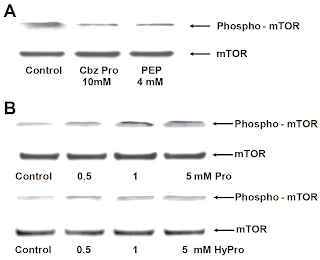
Activated TGF-β1 receptors activate MAPK, AKT and mTOR via phosphorylation. While the prolidase inhibitors had no effect of MAPK, they decreased the expression of phospho-AKT and phosphor-mTOR. Furthermore, adding Pro and HyPro had no effect on MAPK, but increased expression of phosphor-AKT and phosphor-mTOR in a dose-dependent manner.
Figure 4. Western Blot of Phospho-AKT in a medium of confluent human skin fibroblast. In (A), cells are treated with enzyme inhibitors and in (B) cells are treated with different amounts of proline and hydroxyproline.
Figure 5. Western Blot of Phospho-mTOR in a medium of confluent human skin fibroblast. In (A), cells are treated with enzyme inhibitors and in (B) cells are treated with different amounts of proline and hydroxyproline.
Interestingly, the mTOR inhibitor, rapamycin, was found to also decrease prolidase activity in a dose-dependent manner. Thus, the results suggest that Pro and HyPro are modulators for mTOR activation and rapamycin inhibits mTOR activation by decreasing prolidase activity. This relationship would allow mTOR to detect the availability of Pro before it initiates factors to promote collagen synthesis.
Figure 6. Prolidase activity in confluent human skin fibroblast with varying concentration of Rapamycin.
Discussion
While TGF c was mentioned in the title, I did not see it anywhere else in the paper.
Also, they noted that TGF-β3 was not affected in the same conditions, but it was not clear whether they meant that the prolidase products had no effect or if even the enzyme inhibitors had no effect on TGF-β3 expression. The data for TGF-β3 was not shown so it is impossible to tell. Also, no hypothesis was provided as to the difference in results between TGF-β1 and TGF-β3. Thus we don’t know for sure whether this is due to some error in the experimental procedures or a different reason, such as its structural properties.
Finally, they mentioned how prolidase inhibitors decrease the expression of TGF-β1 receptors and signals activated by TGF-β1. However, it is unclear whether the decrease in the expression of the signals is due to the decrease in the number of receptors, which would lead to the activation of fewer signals, or if the prolidase inhibitors act directly on the expression of the signals.


No comments:
Post a Comment